

Contrast-enhanced endoscopic ultrasound
- PMID: 24949350
- PMCID: PMC4062228
- DOI: 10.7178/eus.03.003
Contrast-enhanced endoscopic ultrasound
Abstract
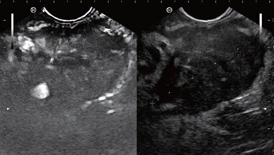
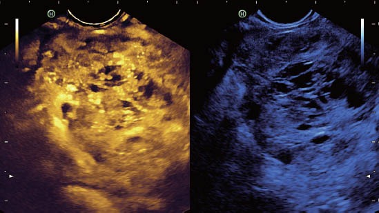
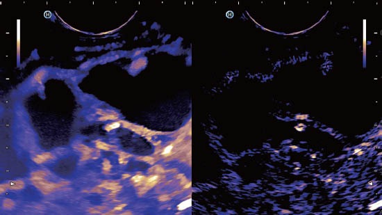
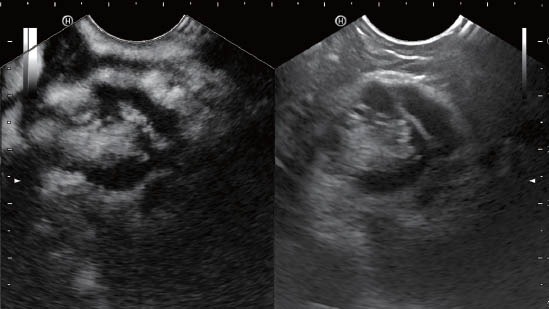
The European Federation of Societies for Ultrasound in Medicine and Biology (EFSUMB) introduced guidelines on the use of contrast-enhanced ultrasound (CEUS) in 2004. This EFSUMB-document focused mainly on liver applications. However, new applications extending beyond the liver were developed thereafter. Increased interest in recent years in CEUS technique and in the application of CEUS in novel fields like endoscopic ultrasound (EUS) has revolutionized indications and applications. As a result, the EFSUMB initiated a new update of the guidelines in 2011 to include this additional knowledge. Some of the contrast-enhanced EUS (CE-EUS) indications are established, whereas others are preliminary; these latter indications are categorized as emergent CEUS applications since the available evidence is insufficient for general recommendation. This article focuses on the use of CE-EUS in various clinical settings. The reader will get an overview of current indications and possible applications of CE-EUS. This involves the introduction of different contrast studies including color Doppler techniques (known as contrast-enhanced high mechanical index endosonography or CEHMI-EUS) as well as more modern high-resolution contrast-enhanced techniques (known as contrast-enhanced low mechanical index endosonography or CELMI EUS).
Keywords: contrast-enhanced; differential diagnosis; endoscopic ultrasound; ultrasound.
Figures
Similar articles
-
EFSUMB guidelines 2011: comments and illustrations.Ultraschall Med. 2012 Jul;33 Suppl 1:S11-21. doi: 10.1055/s-0032-1312890. Epub 2012 Jun 21. Ultraschall Med. 2012. PMID: 22723024 Review.
-
Advanced endosonographic diagnostic tools for discrimination of focal chronic pancreatitis and pancreatic carcinoma--elastography, contrast enhanced high mechanical index (CEHMI) and low mechanical index (CELMI) endosonography in direct comparison.Z Gastroenterol. 2012 Feb;50(2):199-203. doi: 10.1055/s-0031-1281824. Epub 2012 Feb 1. Z Gastroenterol. 2012. PMID: 22298098
-
European Federation of Societies for Ultrasound in Medicine and Biology (EFSUMB): An Update on the Pediatric CEUS Registry on Behalf of the "EFSUMB Pediatric CEUS Registry Working Group".Ultraschall Med. 2021 Jun;42(3):270-277. doi: 10.1055/a-1345-3626. Epub 2021 Mar 9. Ultraschall Med. 2021. PMID: 33690876 English.
-
[Contrast-enhanced endoscopic ultrasound in the diagnosis of pancreatic diseases].Dtsch Med Wochenschr. 2013 Apr;138(14):732-4. doi: 10.1055/s-0032-1332963. Epub 2013 Mar 26. Dtsch Med Wochenschr. 2013. PMID: 23533041 Review. German.
-
The Asian Federation of Societies for Ultrasound in Medicine and Biology (AFSUMB) Guidelines for Contrast-Enhanced Endoscopic Ultrasound.Ultrasound Med Biol. 2021 Jun;47(6):1433-1447. doi: 10.1016/j.ultrasmedbio.2021.01.030. Epub 2021 Feb 27. Ultrasound Med Biol. 2021. PMID: 33653627
Cited by
-
Discriminating chronic pancreatitis from pancreatic cancer: Contrast-enhanced EUS and multidetector computed tomography in direct comparison.Endosc Ultrasound. 2018 Nov-Dec;7(6):395-403. doi: 10.4103/eus.eus_24_18. Endosc Ultrasound. 2018. PMID: 30246709 Free PMC article.
-
Endoscopic ultrasound assessment and tissue acquisition of mediastinal and abdominal lymph nodes.World J Gastrointest Oncol. 2021 Oct 15;13(10):1475-1491. doi: 10.4251/wjgo.v13.i10.1475. World J Gastrointest Oncol. 2021. PMID: 34721779 Free PMC article. Review.
-
Basic Principles and Role of Endoscopic Ultrasound in Diagnosis and Differentiation of Pancreatic Cancer from Other Pancreatic Lesions: A Comprehensive Review of Endoscopic Ultrasound for Pancreatic Cancer.J Clin Med. 2024 Apr 28;13(9):2599. doi: 10.3390/jcm13092599. J Clin Med. 2024. PMID: 38731128 Free PMC article. Review.
-
Endoscopic ultrasonography: an advancing option with duality in both diagnosis and treatment of gastrointestinal oncology.Chin J Cancer Res. 2014 Dec;26(6):724-9. doi: 10.3978/j.issn.1000-9604.2014.12.14. Chin J Cancer Res. 2014. PMID: 25561772 Free PMC article.
-
Dynamic contrast-enhanced endoscopic ultrasound: A quantification method.Endosc Ultrasound. 2017 Jan-Feb;6(1):12-20. doi: 10.4103/2303-9027.193595. Endosc Ultrasound. 2017. PMID: 28218195 Free PMC article. Review.
References
-
- Dietrich CF, Ignee A, Frey H. Contrast-enhanced endoscopic ultrasound with low mechanical index: a new technique. Z Gastroenterol. 2005;43:1219–23. - PubMed
-
- Becker D, Strobel D, Bernatik T, et al. Echo-enhanced color-and power-Doppler EUS for the discrimination between focal pancreatitis and pancreatic carcinoma. Gastrointest Endosc. 2001;53:784–9. - PubMed
-
- Bhutani MS, Hoffman BJ, van Velse A, et al. Contrast-enhanced endoscopic ultrasonography with galactose microparticles: SHU508 A (Levovist) Endoscopy. 1997;29:635–9. - PubMed
-
- Giovannini M. Contrast-enhanced endoscopic ultrasound and elastosonoendoscopy. Best Pract Res Clin Gastroenterol. 2009;23:767–79. - PubMed
-
- Giovannini M. Contrast-enhanced and 3-dimensional endoscopic ultrasonography. Gastroenterol Clin North Am. 2010;39:845–58. - PubMed
Publication types
LinkOut - more resources
Full Text Sources
Research Materials