. 2014:2014:424982.
doi: 10.1155/2014/424982. Epub 2014 May 18.

Circadian modulation of the Cl(-) equilibrium potential in the rat suprachiasmatic nuclei

Affiliations

Circadian modulation of the Cl(-) equilibrium potential in the rat suprachiasmatic nuclei

Javier Alamilla et al. Biomed Res Int. 2014.

Abstract

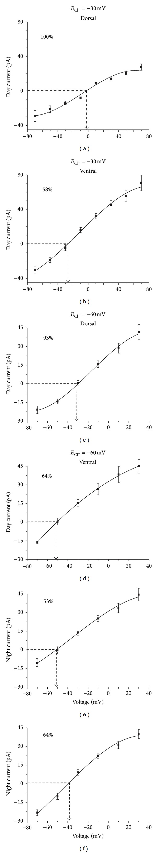
The suprachiasmatic nuclei (SCN) constitute a circadian clock in mammals, where γ-amino-butyric acid (GABA) neurotransmission prevails and participates in different aspects of circadian regulation. Evidence suggests that GABA has an excitatory function in the SCN in addition to its typical inhibitory role. To examine this possibility further, we determined the equilibrium potential of GABAergic postsynaptic currents (E(GABA)) at different times of the day and in different regions of the SCN, using either perforated or whole cell patch clamp. Our results indicate that during the day most neurons in the dorsal SCN have an E(GABA) close to -30 mV while in the ventral SCN they have an E(GABA) close to -60 mV; this difference reverses during the night, in the dorsal SCN neurons have an E(GABA) of -60 mV and in the ventral SCN they have an E(GABA) of -30 mV. The depolarized equilibrium potential can be attributed to the activity of the Na(+)-K(+)-2Cl(-) (NKCC) cotransporter since the equilibrium potential becomes more negative following addition of the NKCC blocker bumetanide. Our results suggest an excitatory role for GABA in the SCN and further indicate both time (day versus night) and regional (dorsal versus ventral) modulation of E(GABA) in the SCN.


Figures

Figure 1
Figure 1
Spontaneous GABAergic postsynaptic currents in the suprachiasmatic nucleus. GABA sPSCs were pharmacologically isolated by administration of the ionotropic glutamate receptor antagonists, DNQX (10 μM) and APV (50 μM). Recordings at 0 mV and −70 mV are shown. Subsequent bicuculline administration (10 μM) completely abolished the sPSCs at all membrane holding potentials. Example recordings obtained in a dorsal SCN neuron during the day period in perforated patch configuration.
Figure 2
Figure 2
Frequency histograms of E GABA obtained using perforated patch recordings. Gaussian fitting analyses revealed two populations in E GABA distribution, belonging to (a) all the neurons recorded in different ZT and SCN regions, (b, c) E GABA sorted by dorsal and ventral SCN regions, and (d, e) E GABA grouped by ZT of recording.
Figure 3
Figure 3
E GABA distributions in the SCN. Comparison of all E GABA values recorded in perforated patch configuration, grouped by SCN region (a) or ZT of recording (b). Values are shown in mean ± SEM. (c) Distribution of E GABA separated by SCN region and ZT of recording. Experiments obtained in perforated patch configuration. (d) Statistically significant differences were found with Kruskal-Wallis test (H = 42.4, P < 0.0001). Analysis was performed with the prevailing part of the groups depicted in (c). Dunn's multiple comparison test indicated significant differences between the groups (P < 0.05). Distribution of E GABA in neurons recorded in whole cell configuration with a theoretical E Cl of −30 mV (e) and E Cl of −60 mV (f).
Figure 4
Figure 4
Diurnal and regional modulation of E GABA from neurons recorded using perforated patch. During the day, the predominant equilibrium potential from the dorsal SCN neurons is −37 ± 2 mV (a) and in the ventral SCN is −61 ± 2 mV (b); during the night, the pattern reverses so that the predominant equilibrium potential from the dorsal SCN is −60 ± 1 mV (c) and in the ventral SCN is −38 ± 2 mV (d). Each panel shows the percentage of neurons contributing to each I-V curve.
Figure 5
Figure 5
E GABA in dorsal SCN region. (a) Perforated patch recording in a dorsal SCN neuron obtained during the day. E GABA for this neuron was −33 mV, as shown in the I-V curve below. (b) Recording performed using perforated patch in a dorsal SCN neuron during the nocturnal ZT. The E GABA for this example was −62 mV (I-V curve below).
Figure 6
Figure 6
E GABA in ventral SCN region. (a) Neuron recorded in perforated patch in a ventral SCN neuron during the day. E GABA for this neuron was −58 mV (I-V curve below). (b) Perforated patch recording performed during the night period in a ventral SCN neuron. E GABA for this cell was −33 mV (I-V curve below).
Figure 7
Figure 7
E GABA is shifted from the E Cl estimated from the Nernst equation in subpopulations of the SCN neurons recorded in whole cell configuration. At an estimated E Cl of −30 mV, the dorsal SCN neurons recorded during the day showed an E GABA of 0 ± 2 mV (a), while in ventral SCN the predominant E GABA is −28 ± 3 mV (b), close to the hypothetical equilibrium potential. At an estimated E Cl of −60 mV, when recorded during day, the predominant GABAergic sPSCs equilibrium potential in the dorsal SCN was −32 ± 2.6 mV (c), while in the ventral SCN was −51 ± 1.4 mV (d). Conversely, during the night, the predominant equilibrium potential in the dorsal SCN was −53 ± 2.3 mV (e) and in the ventral SCN was −39 ± 1.9 mV (f).
Figure 8
Figure 8
Regional differences in E GABA obtained in whole cell configuration. (a) Dorsal SCN neuron recorded during the day in whole cell. E GABA for this neuron was −30 mV (I-V curve below). (b) Ventral SCN neuron recorded during the day in whole cell configuration. E GABA for this neuron was −50 mV (I-V curve below). Theoretical E Cl = −60 mV.
Figure 9
Figure 9
Integrative model of excitatory GABA actions depending on SCN region and circadian time. (a) Dorsal day SCN neurons. High expression of NKCC1 cotransporters increases the [Cl]i. GABAA R activation induces Cl efflux, which depolarizes the E m. Depolarized E m increases the probability of action potentials that strengthens the SFR during the day. (b) Dorsal night. Low expression of NKCC1 decreases the [Cl]i. GABA binding induces Cl influx, which hyperpolarizes the E m. More negative E m decreases the probability of spikes that reduces the SFR during the night. (c) Ventral day. Diminished NKCC1 expression reduces [Cl]i, and GABAA R activation hyperpolarizes E m. Less excitability in ventral neurons decreases the probability of phase shifts. (d) Ventral night. Upregulated NKCC1 expression generates high [Cl]i that shifts the E m close to the spike threshold. More excitability in ventral SCN neurons during the night enhances the probability of phase shifts by GABA.

References

    1. Klein DC, Moore RY, Reppert SM. Suprachiasmatic Nucleus: The Mind's Clock. New York, NY, USA: Oxford University Press; 1991.
    1. Abrahamson EE, Moore RY. Suprachiasmatic nucleus in the mouse: retinal innervation, intrinsic organization and efferent projections. Brain Research. 2001;916(1-2):172–191. - PubMed
    1. LeSauter J, Kriegsfeld LJ, Hon J, Silver R. Calbindin-D28K cells selectively contact intra-SCN neurons. Neuroscience. 2002;111(3):575–585. - PMC - PubMed
    1. Morin LP, Shivers K-Y, Blanchard JH, Muscat L. Complex organization of mouse and rat suprachiasmatic nucleus. Neuroscience. 2006;137(4):1285–1297. - PubMed
    1. Reppert SM, Weaver DR. Coordination of circadian timing in mammals. Nature. 2002;418(6901):935–941. - PubMed

Publication types

MeSH terms

LinkOut - more resources