. 2014 Jun 20;9(6):e100264.
doi: 10.1371/journal.pone.0100264. eCollection 2014.

Pathogenicity of Shigella in chickens

Affiliations

Pathogenicity of Shigella in chickens

Run Shi et al. PLoS One. .

Abstract

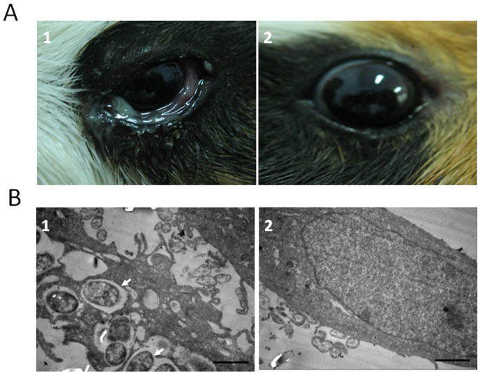
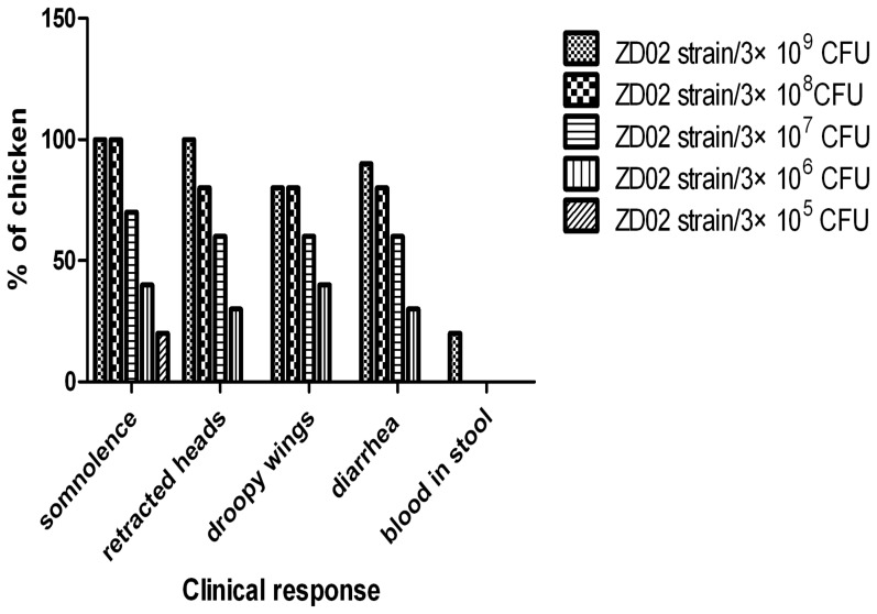
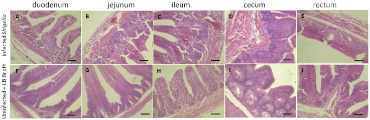
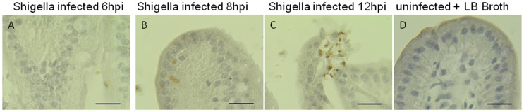
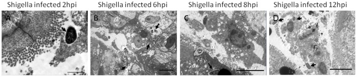
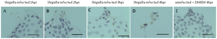
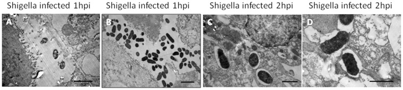
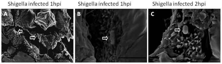
Shigellosis in chickens was first reported in 2004. This study aimed to determine the pathogenicity of Shigella in chickens and the possibility of cross-infection between humans and chickens. The pathogenicity of Shigella in chickens was examined via infection of three-day-old SPF chickens with Shigella strain ZD02 isolated from a human patient. The virulence and invasiveness were examined by infection of the chicken intestines and primary chicken intestinal epithelial cells. The results showed Shigella can cause death via intraperitoneal injection in SPF chickens, but only induce depression via crop injection. Immunohistochemistry and transmission electron microscopy revealed the Shigella can invade the intestinal epithelia. Immunohistochemistry of the primary chicken intestinal epithelial cells infected with Shigella showed the bacteria were internalized into the epithelial cells. Electron microscopy also confirmed that Shigella invaded primary chicken intestinal epithelia and was encapsulated by phagosome-like membranes. Our data demonstrate that Shigella can invade primary chicken intestinal epithelial cells in vitro and chicken intestinal mucosa in vivo, resulting in pathogenicity and even death. The findings suggest Shigella isolated from human or chicken share similar pathogenicity as well as the possibility of human-poultry cross-infection, which is of public health significance.


Conflict of interest statement

Competing Interests: The authors have declared that no competing interests exist.

Figures

Figure 1
Figure 1. The virulence of the human Shigella strain ZD02 using the Sereny test and the HeLa cell invasiveness test.
(A) Sereny test. Guinea pigs were infected with Shigella strain ZD02 via the cornea and conjunctiva. Corneal and conjunctiva inflammation were seen in infected eyes at 18 h post-inoculation (A1). Corneas and conjunctiva is normal in uninfected control eyes (A2). (B) HeLa cells infected with the Shigella strain ZD02. Transmission electron microscopy was used to detect the internalized bacteria within HeLa cells. Representative pictures are shown. The internalized bacteria (arrowheads) were detected at 2 h post-inoculation (B1). Control HeLa cells without infection (B2). Scale bars = 1 µm.
Figure 2
Figure 2. Clinical responses of chickens after intraperitoneal injection.
Each bar denotes number of the clinical response/total in each experimental group after 12 h of intraperitoneal injection with Shigella strain ZD02.
Figure 3
Figure 3. Histopathology of the intestine of one-day-old SPF chickens infected with the Shigella strain ZD02 via ligated intestinal loop.
The duodenum, jejunum, ileum, and cecum from Shigella-infected chickens showed congestion, edema, and neutrophil infiltration (A–E, respectively). No significant pathological changes were noticed in the uninfected controls (F–J, respectively). The sections were stained with hematoxylin and eosin. Representative pictures are shown. Scale bars = 50 µm.
Figure 4
Figure 4. Immunohistochemistry of the jejunum of one-day-old SPF chickens infected with the Shigella strain ZD02 via ligated intestinal loop.
Bacterial invasion (brown staining) was noticed in the epithelial cells 6 h (A) and 8 h (B) post-inoculation. The intestinal villus detached and the bacteria (brown staining) were released from the lysed epithelial cells (C). Intestinal villus of uninfected chicken was shown as control (D). Scale bars = 20 µm.
Figure 5
Figure 5. Transmission electron microscopy of the jejunum of one-day-old SPF chickens infected with the Shigella strain ZD02 via ligated intestinal loop.
Shigella (arrowheads) were found near the epithelial cells at 2 h post-inoculation (A. Scale bar = 1 µm). Internalized bacteria (arrowheads) in the epithelial cells were found at 6 h (B. Scale bar = 5 µm) and 8 h (C. Scale bar = 5 µm). The epithelial cells started to lyse and release bacteria (arrowheads) at 12 h (D. Scale bar = 5 µm).
Figure 6
Figure 6. Immunohistochemistry of the primary chicken intestinal epithelial cells infected with the Shigella strain ZD02.
Uninternalized bacteria (brown staining) were seen near the epithelial cells at 1 h post-inoculation (A). Internalized bacteria (brown staining) were found at 2 h (B) and 3 h (C). The lysed epithelial cells released bacteria (brown staining) at 4 h (D). Uninfected epithelial cells were shown as control (E). Scale bars = 20 µm.
Figure 7
Figure 7. Transmission electron microscopy of the primary chicken intestinal epithelial cells infected with the Shigella strain ZD02.
Uninternalized bacteria were seen near the epithelial cells at 1-inoculation (A, B. Scale bars = 2 µm). The internalized bacteria were encapsulated with phagosome-like membranes (C, D. Scale bars = 1 µm).
Figure 8
Figure 8. Scanning electron microscopy of the primary chicken intestinal epithelial cells infected with the Shigella strain ZD02.
Uninternalized bacteria (arrowhead) were seen near the epithelial cells 1 h post-inoculation (A. Scale bar = 10 µm). Bacteria (arrowheads) attached to the surfaces of the epithelial cells at 1 h (B. Scale bar = 3 µm). Bacteria (arrowheads) were being internalized by the epithelial cells at 2 h (C. Scale bar = 3 µm).

References

    1. Kotloff KL, Winickoff JP, Ivanoff B, Clemens JD, Swerdlow DL, et al. (1999) Global burden of Shigella infections: implications for vaccine development and implementation of control strategies. B World Health Organ 77: 651–666. - PMC - PubMed
    1. Bardhan P, Faruque ASG, Naheed A, Sack DA (2010) Decreasing Shigellosis-related Deaths without Shigella spp.-specific Interventions, Asia. Emerg Infect Dis 16: 1718–1723. - PMC - PubMed
    1. Pan BJ, Wang WL, Xie YP, Wei ML, Luo ZF, et al. (2006) Detection, serology classification and drug susceptibility of Shigella from experimental monkeys. Guangxi Agri Sci 37: 331–332.
    1. Jiang JJ, Wang PY, Pan GQ, Kang LC (2005) Isolation and Identification of Rabbits Shigella Dysenteriae in a Large Scale Warren. J Anhui Agri Sci 33: 1666–1667.
    1. Priamukhima NS, Kilesso VA, Tikhomirov ED (1984) Animal carriers of Shigella and their possible epidemiological importance. Zh Mikrobiol Epidemiol Immunobiol 11: 20–24. - PubMed

Publication types

LinkOut - more resources