. 2014 Sep;155(9):3227-37.
doi: 10.1210/en.2014-1016. Epub 2014 Jun 20.

The vitamin D receptor (VDR) is expressed in skeletal muscle of male mice and modulates 25-hydroxyvitamin D (25OHD) uptake in myofibers

Affiliations

The vitamin D receptor (VDR) is expressed in skeletal muscle of male mice and modulates 25-hydroxyvitamin D (25OHD) uptake in myofibers

Christian M Girgis et al. Endocrinology. 2014 Sep.

Abstract

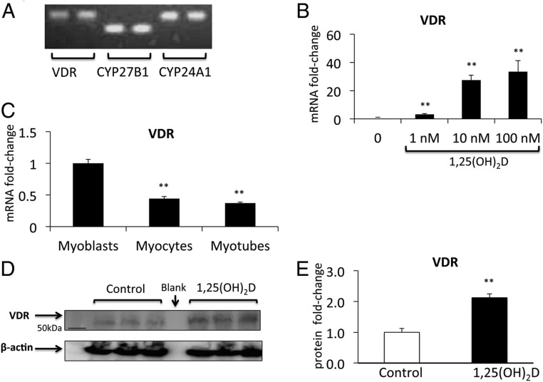
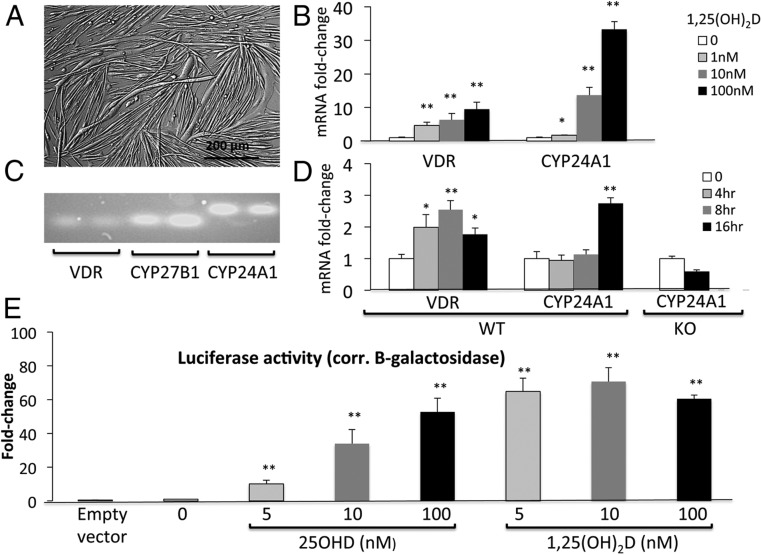
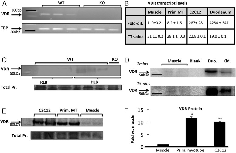
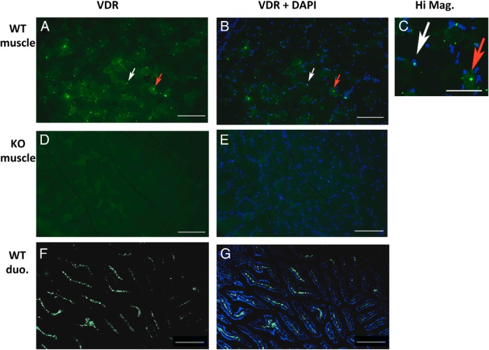
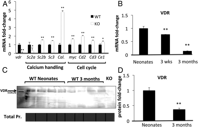
Vitamin D deficiency is associated with a range of muscle disorders, including myalgia, muscle weakness, and falls. In humans, polymorphisms of the vitamin D receptor (VDR) gene are associated with variations in muscle strength, and in mice, genetic ablation of VDR results in muscle fiber atrophy and motor deficits. However, mechanisms by which VDR regulates muscle function and morphology remain unclear. A crucial question is whether VDR is expressed in skeletal muscle and directly alters muscle physiology. Using PCR, Western blotting, and immunohistochemistry (VDR-D6 antibody), we detected VDR in murine quadriceps muscle. Detection by Western blotting was dependent on the use of hyperosmolar lysis buffer. Levels of VDR in muscle were low compared with duodenum and dropped progressively with age. Two in vitro models, C2C12 and primary myotubes, displayed dose- and time-dependent increases in expression of both VDR and its target gene CYP24A1 after 1,25(OH)2D (1,25 dihydroxyvitamin D) treatment. Primary myotubes also expressed functional CYP27B1 as demonstrated by luciferase reporter studies, supporting an autoregulatory vitamin D-endocrine system in muscle. Myofibers isolated from mice retained tritiated 25-hydroxyvitamin D3, and this increased after 3 hours of pretreatment with 1,25(OH)2D (0.1 nM). No such response was seen in myofibers from VDR knockout mice. In summary, VDR is expressed in skeletal muscle, and vitamin D regulates gene expression and modulates ligand-dependent uptake of 25-hydroxyvitamin D3 in primary myofibers.


Figures

Figure 1.
Figure 1.
Components of the vitamin D-endocrine system in C2C12 muscle cells. A, C2C12 cells express CYP27B1, VDR, and CYP24A1 mRNA as seen on semiquantitative PCR. Duplicates for each have been shown. B, Expression of VDR mRNA is stimulated in a dose-dependent fashion by 48 hours of treatment with 1,25(OH)2D (data are mean ± SEM, n = 3 per group). C, VDR mRNA drops sequentially during C2C12 differentiation from myoblasts to myotubes (P < .005). Western blotting (D) and densitometric quantitation (E) show that VDR expression (normalized for β-actin) increased 2.2-fold in response to 72 hours of treatment with 1,25(OH)2D in C2C12 myotubes (P < .005, n = 3 per group).
Figure 2.
Figure 2.
Components of the vitamin D-endocrine system in primary myotubes. A, Primary myotubes are elongated, multinucleated syncytia that contract in culture and resemble muscle fibers. B, On RT-PCR, expression of VDR and CYP24A1 mRNA is stimulated in a dose-dependent fashion by 48 hours of treatment with 1,25(OH)2D (mean ± SEM, n = 3 per group). C, Primary myotubes express CYP27B1, VDR, and CYP24A1 mRNA as seen on semiquantitative PCR. Duplicates for each have been shown. D, On RT-PCR, expression of VDR increases in a time-dependent fashion after treatment with 100nM 1,25(OH)2D. An increase in CYP24A1 was first noted at 16 hours in myotubes from WT mice, but this effect was absent in myotubes from KO mice. E, 25OHD induced a dose-dependent increase in luciferase activity in primary myotubes transfected with Gal4-VDR (switch) and UASTK-luciferase reporter (P < .005, n = 4–6 per group). Similar changes were observed in response to 1,25(OH)2D (P < .005, n = 4–6 per group). Luciferase activity was corrected for β-galactosidase activity as transfection control.
Figure 3.
Figure 3.
Detection of VDR in skeletal muscle on PCR and Western blotting. A, On semiquantitative PCR, VDR transcript is detectable in quadriceps muscle from 3 adult WT mice. Muscle from 3 VDRKO mice was used as negative control. B, VDR transcript levels relative to whole muscle (ie, fold difference; VDR mRNA levels divided by that in whole muscle) and absolute CT values are listed (mean ± SEM, n = 3 per group). VDR transcript levels are markedly higher in duodenum (Duo.), the classic target of vitamin D action, compared with whole muscle and appreciably higher in muscle cell models compared with whole muscle. C, Using VDR-D6 antibody on Western blotting, VDR was detected in quadriceps muscle processed in HLB but hardly detected in samples processed in RLB (n = 9 per group, 60-μg protein/well, 7.2-fold higher detection; P < .05). For negative control, VDRKO muscle sample processed in HLB was used (n = 3). D, Compared with Duo. and kidney (Kid.), VDR in muscle required longer exposure time (15 min) and more protein loading (50 vs 10 μg per lane). Western blotting (E) and ImageJ densitometric quantitation (F) show that VDR expression (normalized for total protein on Coomassie) is approximately 10-fold higher in primary myotubes (Prim. MT) and C2C12 myotubes compared with whole muscle (n = 2 per group, P < .005, 20-μg protein/well). Samples used were processed in HLB. TBP, TATA box binding protein.
Figure 4.
Figure 4.
Detection of VDR in skeletal muscle on immunohistochemistry. Using VDR-D6 antibody, VDR was detected within muscle fibers of young WT mice on immunohistochemistry (8-μm cross-sections, quadriceps) (A) and localized to fiber nuclei (white arrow) and cytoplasm (red arrow) as seen on DAPI counterstaining (scale bars, 100 μm) (B). C, This is further demonstrated on higher magnification (scale bar, 50 μm; image brightness adjusted to aid in visualizing the cytoplasm). D and E, No such signal was detected in muscle from age-matched VDRKO mice in which DAPI staining was also performed. F and G, Duodenum (duo.) from adult WT mice was used as positive control and showed substantially greater levels of VDR than muscle (scale bars, 100 μm).
Figure 5.
Figure 5.
Effects of VDRKO in muscle gene expression and age-related differences in muscle VDR in WT mice. A, Muscle from WT vs VDRKO mice displayed significant differences in the mRNAs of calcium-handling genes, including Serca2a, Serca2b, Serca3, and Calbindin-28K (Sc2a, Sc2b, Sc3, and Cal.), and cell cycle regulatory genes, including myc and Cyclins D2, D3, and E1 (Cd2, Cd3, and Ce1). B, VDR transcript levels were significantly higher in muscles of newborn WT mice compared with 3-week- and 3-month-old mice (n = 3 per group; P < .005). Using samples prepared in HLB, Western blotting (C) and densitometric quantitation (D) demonstrated higher VDR expression in muscles of newborn WT mice compared with 3-month-old mice (n = 4 per group; P < .005). Muscle from newborn VDRKO mice was used as negative control. Pr, protein.
Figure 6.
Figure 6.
3H-25OHD3 uptake in WT and VDRKO myofibers. A, Preincubation of WT myofibers with 1,25(OH)2D resulted in 40% greater 3H-25OHD3 uptake compared with WT myofibers preincubated with control solution (P < .005). No such increase in 3H-25OHD3 uptake in response to 1,25(OH)2D was seen in VDRKO mice (n = 3 mice per group). B, In C2C12 myotubes, DIDS reversed the 1,25(OH)2D-mediated increase in uptake of 3H-25OHD3. cpm, counts per minute.

Comment in

References

    1. Glisson F. A Treatise of the Rickets: Being a Disease Common to Children. Culpeper N, ed. London, UK: P. Cole; 1651.
    1. Girgis CM, Clifton-Bligh RJ, Hamrick MW, Holick MF, Gunton JE. The roles of vitamin D in skeletal muscle: form, function, and metabolism. Endocr Rev. 2013;34:33–83. - PubMed
    1. Murad MH, Elamin KB, Abu Elnour NO, et al. The effect of vitamin D on falls: a systematic review and meta-analysis. J Clin Endocrinol Metab. 2011;96:1911–1930. - PubMed
    1. Ward KA, Das G, Roberts SA, et al. A randomized, controlled trial of vitamin D supplementation upon musculoskeletal health in postmenarchal females. J Clin Endocrinol Metab. 2010;95:4643–4651. - PubMed
    1. Sohl E, van Schoor NM, de Jongh RT, Visser M, Deeg DJ, Lips P. Vitamin d status is associated with functional limitations and functional decline in older individuals. J Clin Endocrinol Metab. 2013;98:E1483–E1490. - PubMed

Publication types