

Delayed patchy choroidal filling in the Comparison of Age-Related Macular Degeneration Treatments Trials (CATT)
- PMID: 24949820
- PMCID: PMC4458330
- DOI: 10.1016/j.ajo.2014.06.004
Delayed patchy choroidal filling in the Comparison of Age-Related Macular Degeneration Treatments Trials (CATT)
Abstract
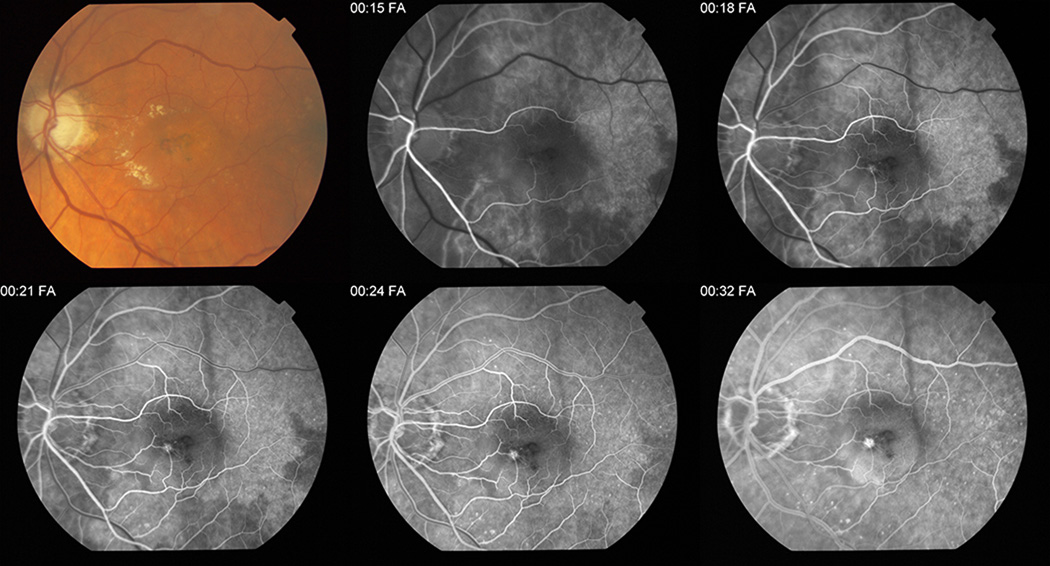
Purpose: To determine the relationship between delayed patchy choroidal filling and morphologic and functional outcomes among eyes treated with ranibizumab or bevacizumab.
Design: Cohort study.
Methods: Comparison of Age-related Macular Degeneration Treatment Trials participants were assigned randomly to ranibizumab or bevacizumab on a monthly or as-needed schedule. Presence of delayed patchy choroidal filling and morphologic and functional outcomes were evaluated among eyes with gradable fluorescein angiography at baseline (n = 973) and at 1 year (n = 860) eyes.
Results: Delayed filling was present in 75 (7.7%) of 973 eyes at baseline. Eyes with incident delayed filling at 1 year (23 [2.9%] of 798) showed a mean decrease of 1.7 letters in visual acuity, whereas eyes without incident delayed filling had a mean improvement of 8.1 letters (difference [Δ], -9.8; 95% confidence interval [CI] , -15.8 to -3.9; P < .01). Eyes with incident delayed filling had a larger increase in mean total lesion area of choroidal neovascularization (3.00 mm(2)) than eyes without incident delayed filling (0.56 mm(2); Δ , 2.4; 95% CI, 0.4 to 4.4; P = .02). The proportion with incident delayed filling at 1 year was similar among eyes treated with ranibizumab (10 [2.4%] of 413) or bevacizumab (13 [3.3%] of 385; P = .53) and among eyes treated monthly (12 [3.1%] of 388) or as needed (11 [2.7%] of 410; P = .83).
Conclusions: Delayed patchy choroidal filling was uncommon at baseline. Although only a small percentage of eyes demonstrated delayed filling during the first year of anti-vascular endothelial growth factor treatment, these eyes had worse visual acuity and a larger increase in total lesion area of choroidal neovascularization.
Copyright © 2014 Elsevier Inc. All rights reserved.
Figures
References
-
- Boltz A, Luksch A, Wimpissinger B, et al. Choroidal blood flow and progression of age-related macular degeneration in the fellow eye in patients with unilateral choroidal neovascularization. Invest Ophthalmol Vis Sci. 2010;51(8):4220–4225. - PubMed
-
- Hayashi K, de Laey JJ. Indocyanine green angiography of choroidal neovascular membranes. Ophthalmologica. 1985;190(1):30–39. - PubMed
-
- Mendrinos E, Pournaras CJ. Topographic variation of the choroidal watershed zone and its relationship to neovascularization in patients with age-related macular degeneration. Acta Ophthalmol. 2009;87(3):290–296. - PubMed
Publication types
MeSH terms
Substances
Grants and funding
LinkOut - more resources
Full Text Sources
Other Literature Sources
Medical
