. 2014 Aug 15;392(2):168-81.
doi: 10.1016/j.ydbio.2014.06.006. Epub 2014 Jun 17.

Multiple tissue-specific requirements for the BMP antagonist Noggin in development of the mammalian craniofacial skeleton

Affiliations

Multiple tissue-specific requirements for the BMP antagonist Noggin in development of the mammalian craniofacial skeleton

Maiko Matsui et al. Dev Biol. .

Abstract

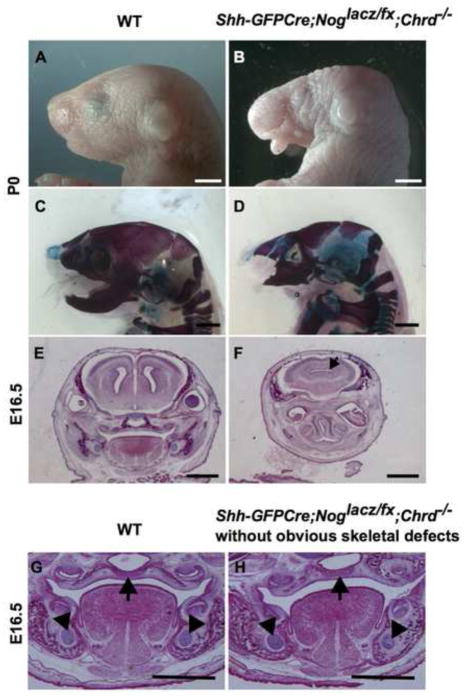
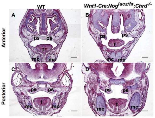
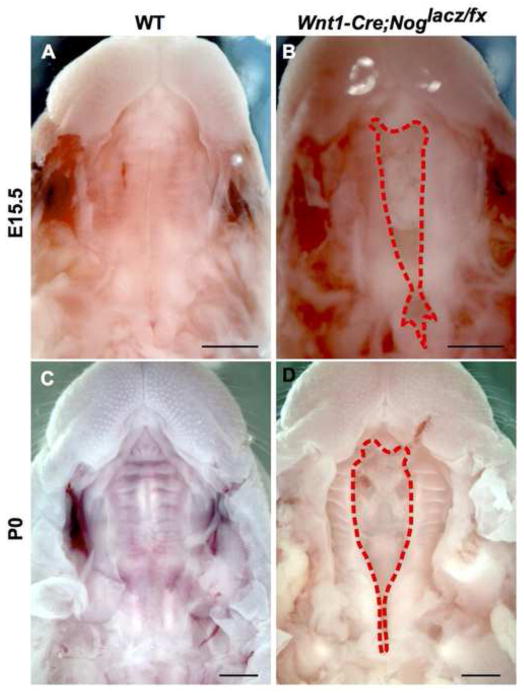
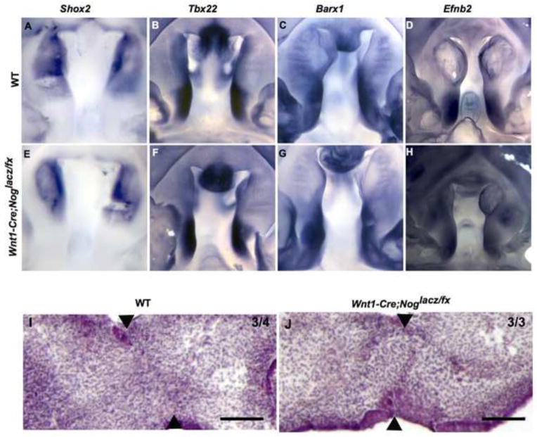
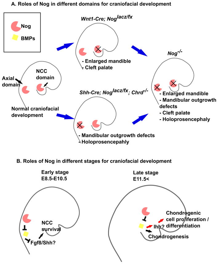
Proper morphogenesis is essential for both form and function of the mammalian craniofacial skeleton, which consists of more than twenty small cartilages and bones. Skeletal elements that support the oral cavity are derived from cranial neural crest cells (NCCs) that develop in the maxillary and mandibular buds of pharyngeal arch 1 (PA1). Bone Morphogenetic Protein (BMP) signaling has been implicated in most aspects of craniofacial skeletogenesis, including PA1 development. However, the roles of the BMP antagonist Noggin in formation of the craniofacial skeleton remain unclear, in part because of its multiple domains of expression during formative stages. Here we used a tissue-specific gene ablation approach to assess roles of Noggin (Nog) in two different tissue domains potentially relevant to mandibular and maxillary development. We found that the axial midline domain of Nog expression is critical to promote PA1 development in early stages, necessary for adequate outgrowth of the mandibular bud. Subsequently, Nog expression in NCCs regulates craniofacial cartilage and bone formation. Mice lacking Nog in NCCs have an enlarged mandible that results from increased cell proliferation in and around Meckel׳s cartilage. These mutants also show complete secondary cleft palate, most likely due to inhibition of posterior palatal shelf elevation by disrupted morphology of the developing skull base. Our findings demonstrate multiple roles of Noggin in different domains for craniofacial skeletogenesis, and suggest an indirect mechanism for secondary cleft palate in Nog mutants that may be relevant to human cleft palate as well.

Keywords: Bone morphogenetic protein; Mandible; Mouse development; Neural crest; Noggin; Palate.


Figures

Fig. 1
Fig. 1. Nog is expressed in multiple domains during craniofacial development
(A, B) Nog is first expressed in migrating NCCs around E8.0. (C) Nog is expressed in the NCCs of PA1. (D) By E10.5 Nog expression is restricted to the oral epithelium of PA1. (E, F) Sections through PA1 showing transient expression of Nog. At E9.0, Nog expression is found in mesenchyme of PA1 (E), but Nog expression subsequently shifts to the epithelium layer of PA1 (F). (G, H) Nog is expressed in the cartilages of the head. pa1; pharyngeal arch 1, fp; floor plate of the neural tube, mc; Meckel’s cartilage, nccs; neural crest cells, ns; nasal septum, oe; oral epithelium.
Fig. 2
Fig. 2. BMP antagonism in axial domain is critical for development of PA1 derivatives
(A–D) Lateral view of wild-type (A, C) and Shh-GFPCre;Noglacz/fx;Chrd−/− (B, D) mutant whole-mount and skeletal-preparation heads at P0 stage showing that a lack of Chrd and Nog in the Shh-GFPCre domain results in hypomorphic PA1 derivatives compared with wild-type littermates. Scale bars = 2 mm. (E, F) Example of holoprosencephaly in mutant embryo at E16.5 (F), with a normal littermate (E). (G, H) Mutant embryos without obvious phenotypes showing relatively normal development of the palate (indicated with an arrow) and Meckel’s cartilage (indicated with arrowheads (H)). Scale bars = 1 mm.
Fig. 3
Fig. 3. Absence of Nog in NCCs causes craniofacial defects
(A, C) Anterior and posterior coronal sections through E15.5 wild-type embryonic head showing fusion of the palatal shelves and normal development of cartilages in the head. (B, D) Anterior and posterior coronal sections of Wnt1-Cre;Noglacz/fx;Chrd−/− mutant embryos showing cleft palate and enlarged cartilages at E15.5. ps; palatal shelves, t; tongue, mc; Meckel’s cartilage. Scale bars = 400 μm.
Fig. 4
Fig. 4. An enlarged Meckel’s cartilage with increased cell proliferation results from upregulation of BMP signaling in neural crest derivatives
(A, B) Mandibular skeletal preparations. Wild type mandible (A) shows normal development of Meckel’s cartilage (blue) and mandibular bone (purple). Double headed arrow shows antero-posterior (a-p) direction of mandibular outgrowth. Enlarged Meckel’s cartilage in Wnt1-Cre;Noglacz/fx mutant (B) shows no obvious outgrowth defects. Scale bar = 1 mm. (C, D) Meckel’s cartilage surrounded by a red dotted line. The size of wild type (C) Meckel’s cartilage is similar to mutant (D) at E11.5. Scale bars = 100 μm. (E, F) Cell proliferation in Meckel’s cartilage as indicated by BrdU incorporation. Compared with cell proliferation in wild-type Meckel’s cartilage (E), cell proliferation was increased in the mutant Meckel’s cartilage at E12.5 (F). Scale bars = 100 μm. (G, H) Meckel’s cartilage surrounded by a red dotted line. Expression of a constitutively active BMP receptor 1a (caBMPr1a) in combination with Wnt1-Cre caused ectopic expression of Hh signaling in mutant Meckel’s cartilage, as revealed by blue staining of Ptch-lacz activity at E13.5 (H). Wild-type (G) Meckel’s cartilage showed no beta-galactosidase activity. Scale bar = 100 μm. (I) Mandibular outgrowth in the antero-posterior direction as shown in (A) is not different between wild-type and mutant embryos. (J) The diameter of Meckel’s cartilage showed similar size between wild-type and mutant at E11.5. However, after E12.5 mutant Meckel’s cartilage started rapidly growing and the growth persisted at E15.5, while growth rate in wild-type Meckel’s cartilage was diminished. (K) Increased cell proliferation in both perichondrium (peri) and mesenchyme (mes) in Meckel’s cartilage of Wnt1-Cre;Noglacz/fx mutant was statistically significant at E12.5. (L) qPCR indicates increased expression of Hh signaling targets (Gli1, Ptch1) and the Ihh ligand gene in the mutant Meckel’s cartilage at E12.5. P<0.05.
Fig. 5
Fig. 5. Nog in NCCs is required for proper palatogenesis
(A, B) Ventral views of palatal structure. A wild-type E15.5 mouse (A) showing normal palatal development in which the palatal shelves have fused by this stage, compared with the secondary cleft palate in a Wnt1-Cre;Noglacz/fx embryo (B). (C, D) Ventral views of palatal structure in newborn pups (P0). A wild-type P0 mouse showing normal palatal development (C). Overt cleft palate in Wnt1-Cre;Noglacz/fx is obvious at P0 (D). Scale bars = 1 mm.
Fig. 6
Fig. 6. Palatal shelf development in Wnt1-Cre;Noglacz/fx appears normal before elevation
(A–F) Anterior to posterior level matched coronal sections of E13.5 control (A–C) and mutant (D–F) littermates. (G– L) Anterior to posterior level matched coronal sections of E14.5 control (G–I) and mutant (J–L) littermates. Sections shown in the left column (A, D) are anterior secondary palate. Sections shown in the left column (G, J) are from the elevated secondary palate region. Mutant palatal shelves (J) often show incomplete elevation of palatal shelves. Sections shown in the middle column (B, E, H, K) are from the middle of the secondary palate region. Sections shown in the right column (C, F, I, L) are from the posterior soft palate region. (M, N) numbers of BrdU-positive cells per unit area in the palatal shelves of E12.5 (M) and E13.5 (N) were counted and compared between wild-type and Wnt1-Cre;Noglacz/fx mutant littermates. They showed no statistically significant difference in cell proliferation. ns, nasal septum; ps, palatal shelf. Scale bars = 400 μm.
Fig. 7
Fig. 7. Lack of Nog in NCCs does not prevent normal patterning or fusion steps of palatal development
(A–E) Anterior and posterior local patterning characteristics in the secondary palatal shelves are not altered in the Wnt1-Cre;Noglacz/fx mutant mice. (A, E) Shox2 mRNA expression was restricted to the anterior palatal shelves in E13.5 wild-type (A) and mutant (E) littermates. (B–D, F–H) Expression of Tbx22 (B, F), Barx1 (C, G), and Efnb2 (D, H) mRNA was restricted to the posterior region of the E13.5 palatal shelves in both wild-type (B–D) and mutant (F–H) embryos. (I, J) Histological sections of cultured wildtype (I) and Wnt1-Cre;Noglacz/fx mutant (J) palatal shelves juxtaposed in culture show that both genotypes can fuse normally. Arrowheads indicate the point where palatal shelf fusion has taken place. Scale bars = 100 μm.
Fig. 8
Fig. 8. Pterygoid bone disrupts the posterior palatal shelves in the Wnt1-Cre;Noglacz/fx mutant palate
(A–F) Coronal sections of heads from E13.5 and E14.5, comparing palatal structure in wild type and Wnt1-Cre;Noglacz/fx mutants. Before palatal shelf elevation at E13.5, the pterygoid bone is not obvious in this plane of section in the wild-type embryo (A), but it is already much larger in the mutant and invades into the posterior part of palatal shelves (B). (C, D) Coronal sections through posterior palate at E14.5. The pterygoid bone appears in wild-type at E14.5 (C), while it intrudes much further into mutant palatal shelves at this stage (D). (E, F) Coronal sections of palatal bone region at E14.5. Completely elevated and fused wild-type palatal shelves (E), whereas palatal shelves were elevated but the angle of elevation was wider in the Wnt1-Cre;Noglacz/fx mutant littermate (F). (G) The angle of palatal elevation was recorded and compared between wild-type and mutant littermates. (H, I) Skeletal preparation showing ventral view of the posterior skull base and the palate. Compared with the width of wild-type skull (H), the width of mutant skull is wider (I). (J, K) Isolated skull base and the palatal bone region from the skeletal preparations at E17.5. The bilateral palatal bones are in contact in wild-type embryo (J). In the mutant (K), the palatal bones are smaller and far apart, the pterygoid bone is enlarged and dysmorphic. (L) The length and width of the skull base were recorded and compared between wild-type and mutant skeletal preparations at E17.5. pb, palatal bone; pt, pterygoid bone; prs, presphenoid; bs, basisphenoid; a, anterior; p, posterior; ml, medial-lateral. *P-value < 0.05, **P-value < 0.01. Scale bars = 100 μm for A, B, C, D, 500 μm for E, F.
Fig. 9
Fig. 9. Summary and model of the roles of Nog in development of craniofacial structures
(A) Nog is expressed in two relevant domains for craniofacial development – NCCs and axial midline. When Nog is ablated in NCCs, an enlarged mandible and cleft palate result without mandibular outgrowth defects. When the axial midline domain of Nog is absent, as well as Chordin, encoding a second midline BMP antagonist, mandibular outgrowth defects and holoprosencephaly occur. Nog null mutant embryos exhibit phenotypes of both of these tissue-specific mutants combined: enlarged mandible, cleft palate, mandibular outgrowth defects, and holoprosencephaly. (B) Earlier expression of Nog in axial domains promotes NCC survival by promoting secondary cues such as Fgf8 and Hh signaling. However, at later stages, Nog regulates skeletogenesis by balancing the level of BMP signaling in and around NCC derivatives. In chondrocytes, Nog regulates the size of cartilage by suppressing cell proliferation. When BMP attenuation is insufficient, chondrocyte proliferation is promoted, possibly through ectopic Ihh expression and Hh pathway signaling.

Similar articles

Cited by

References

    1. Anderson RM, Lawrence AR, Stottmann RW, Bachiller D, Klingensmith J. Chordin and noggin promote organizing centers of forebrain development in the mouse. Development. 2002;129:4975–4987. - PubMed
    1. Anderson RM, Stottmann RW, Choi M, Klingensmith J. Endogenous bone morphogenetic protein antagonists regulate mammalian neural crest generation and survival. Dev Dyn. 2006;235:2507–2520. - PMC - PubMed
    1. Bachiller D, Klingensmith J, Kemp C, Belo JA, Anderson RM, May SR, McMahon JA, McMahon AP, Harland RM, Rossant J, De Robertis EM. The organizer factors Chordin and Noggin are required for mouse forebrain development. Nature. 2000;403:658–661. - PubMed
    1. Baek JA, Lan Y, Liu H, Maltby KM, Mishina Y, Jiang RL. Bmpr1a signaling plays critical roles in palatal shelf growth and palatal bone formation. Developmental Biology. 2011;350:520–531. - PMC - PubMed
    1. Belo JA, Bouwmeester T, Leyns L, Kertesz N, Gallo M, Follettie M, De Robertis EM. Cerberus-like is a secreted factor with neutralizing activity expressed in the anterior primitive endoderm of the mouse gastrula. Mech Dev. 1997;68:45–57. - PubMed

Publication types

MeSH terms

LinkOut - more resources