. 2014 Jun 20;9(6):e100513.
doi: 10.1371/journal.pone.0100513. eCollection 2014.

Skeletal muscle expression of the adhesion-GPCR CD97: CD97 deletion induces an abnormal structure of the sarcoplasmatic reticulum but does not impair skeletal muscle function

Affiliations

Skeletal muscle expression of the adhesion-GPCR CD97: CD97 deletion induces an abnormal structure of the sarcoplasmatic reticulum but does not impair skeletal muscle function

Tatiana Zyryanova et al. PLoS One. .

Abstract

CD97 is a widely expressed adhesion class G-protein-coupled receptor (aGPCR). Here, we investigated the presence of CD97 in normal and malignant human skeletal muscle as well as the ultrastructural and functional consequences of CD97 deficiency in mice. In normal human skeletal muscle, CD97 was expressed at the peripheral sarcolemma of all myofibers, as revealed by immunostaining of tissue sections and surface labeling of single myocytes using flow cytometry. In muscle cross-sections, an intracellular polygonal, honeycomb-like CD97-staining pattern, typical for molecules located in the T-tubule or sarcoplasmatic reticulum (SR), was additionally found. CD97 co-localized with SR Ca2+-ATPase (SERCA), a constituent of the longitudinal SR, but not with the receptors for dihydropyridine (DHPR) or ryanodine (RYR), located in the T-tubule and terminal SR, respectively. Intracellular expression of CD97 was higher in slow-twitch compared to most fast-twitch myofibers. In rhabdomyosarcomas, CD97 was strongly upregulated and in part more N-glycosylated compared to normal skeletal muscle. All tumors were strongly CD97-positive, independent of the underlying histological subtype, suggesting high sensitivity of CD97 for this tumor. Ultrastructural analysis of murine skeletal myofibers confirmed the location of CD97 in the SR. CD97 knock-out mice had a dilated SR, resulting in a partial increase in triad diameter yet not affecting the T-tubule, sarcomeric, and mitochondrial structure. Despite these obvious ultrastructural changes, intracellular Ca2+ release from single myofibers, force generation and fatigability of isolated soleus muscles, and wheel-running capacity of mice were not affected by the lack of CD97. We conclude that CD97 is located in the SR and at the peripheral sarcolemma of human and murine skeletal muscle, where its absence affects the structure of the SR without impairing skeletal muscle function.


Conflict of interest statement

Competing Interests: The authors have declared that no competing interests exist.

Figures

Figure 1
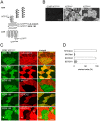
Figure 1. CD97 is expressed in human skeletal muscle.
A Schematic presentation of the human CD97 isoforms containing three (EGF125), four (EGF1235), or five (EGF1-5) EGF-like domains in the NTF . Indicated are the binding sites of the Abs used in this study. The exact binding site of all NTFGAIN Abs within the large GAIN-domain is unknown. B The CD97 NTFEGF1 Ab BL-Ac(F2), detecting an N-glycosylation-dependent epitope in the first EGF-like domains, did not bind to normal human skeletal muscle. In contrast, the sarcolemma of all fibers was stained by the NTFGAIN Ab CLB-CD97/3. The NTFGAIN Abs CLB-CD97/3 and MEM-180, binding to different epitopes within the GAIN domain, intensively stained a subpopulation of muscle fibers intracellularly. C Double immunofluorescence staining of a representative vastus medialis muscle. The fibers that were stained more strongly by CLB-CD97/3 and MEM-180 were the same. Co-staining with the MEM-180 and MHC- or SERCA-specific Abs indicated that slow-twitch fibers were more strongly CD97-positive. A small subpopulation of SERCA1-positive myofibers also stained more strongly for CD97 (asterisk). D Percentage of MEM-180 strongly-positive fibers, set to 100%, co-stained with MHC- and SERCA-specific Abs (n = 170 fibers, mean ± SEM).
Figure 2
Figure 2. CD97 co-localizes with SERCA.
A In cross-sections, CD97 NTFGAIN CLB-CD97/3 and SERCA2 Abs showed a honeycomb-like staining pattern, indicating localization of CD97 within the SR or T-tubules (scale bar 10 µm). A blurred, fuzzy staining was seen with the CD97 NTFGAIN Ab MEM-180 inside the fibers. B In longitudinal sections, a striated pattern was seen with CLB-CD97/3 and MEM-180, whereas staining for RYR and DHPR yielded two parallel dotted lines. C The CD97 Abs CLB-CD97/3 and NTFEndo3 ab13345 co-localized with SERCA2 in longitudinal sections, whereas the MEM-180 projected to the M-band.
Figure 3
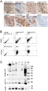
Figure 3. Rhabomyosarcoma strongly express CD97.
A Immunostaining of a rhabomyosarcoma (patient no. 1, table 3; alveolar subtype). The tumor is positive for myogenin and muscle actin (HHF35) but negative for smooth muscle actin (SMA), typical for most rhabdomyosarcomas. Tumor cells were strongly stained by the NTFGAIN CLB-CD97/3 and NTFEndo3 ab13345 Abs. The NTFEGF1 Ab BL-Ac(F2) showed weak staining, indicating partial N-glycosylation of the CD97 EGF-like domains. The NTFGAIN Ab MEM-180 did not stain tumor cells. The tumor infiltrated a smooth muscle which is SMA-positive (arrow) and expresses slightly CD97. Scale bar 50 µm. B In flow cytometry, normal human skeletal muscle cells (SkMC) and Hs729.T cells were CLB-CD97/3-positive, whereas the NTFEGF1 Ab BL-Ac(F2) only stained the tumor cells more strongly, indicating the presence of N-glycosylated CD97. C Western blot analysis using the CD97 NTFGAIN Ab HPA013707 and a CFTICD Ab. After blotting, the membrane was cut horizontally at a height of 34 kDa. The upper part of the blot was incubated with the CD97 NTFGAIN Ab and the lower part with the CFTICD Ab. 1: rhabdomyosarcoma, 2: cultured normal human skeletal muscle cells (SkMC), 3: HS729T cells, 4: duodenum of Tg(CD97-villin) mice , 5: human peripheral blood lymphoytes, 6: human HT1080 cells overexpressing hCD97(EGF125) . Mr, molecular weight marker. The band of about 50 kDa (arrow head) probably represents the naked protein core of the NTF; it is not present in HS729T and peripheral blood lymphocytes. The bands of 70-98 kDa may represent different hCD97 isoforms or differently glycosylated forms. The rhabdomyosarcoma showed an additional high-molecular weight band (asterisk). With the CTFICD Ab, a 25-kDa band representing the CTF was detected in all lysates (arrow) including the murine tissue because this Ab detects human as well as murine CD97. Incubating the entire blot with the CTFICD Ab showed the additional high-molecular weight band in the rhabdomyosarcoma which indicates an uncleaved CD97 form in this tumor (data not shown). In lanes 1-3 and 5, 10 µg, in lane 4, 5 µg, and in lane 6, 2.5 µg protein were separated. Loading was controlled with an α-tubulin Ab, shown in the lower panel.
Figure 4
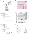
Figure 4. CD97 is present in murine skeletal muscle.
A Schematic presentation of the murine CD97 isoforms containing three (EGF124), four (EGF1234), or four EGF-like domains plus 45 additional amino acids (EGF12×34) . Indicated are the binding sites of the Abs used in this study. The CD97 NTF Ab AF3734 is not shown because its exact binding site within the NTF is unknown. B Nuclear β-galactosidase staining (blue) with fast red counterstaining in the soleus muscle of a CD97-lacZ knock-in mouse; scale bar 50 µm. C, D CD97 mRNA levels of murine tissues (C) and cell lines (D) were quantified by RT-PCR. RSP29 normalized log2 x-fold mRNA levels compared to brain and CMT-93 cells, which were set to one, are given (n = 3, mean ± SEM). E The upper part of the Western blot was incubated with the CD97 NTF Ab AF3734, whereas the lower part was incubated with a CD97 CFTICD Ab. 1: WT mouse, soleus muscle, 2: CD97Ko mouse, soleus muscle, 3: WT mouse, enriched sarcolemma, 4: C2C12 myocytes cultured in 10% FCS, 5: C2C12 myoblasts cultured in 2% horse serum, 6: positive control duodenum Tg(villin-CD97) mouse overexpressing CD97(EGF1234) in intestinal epithelial cells . In lanes 1–2, 20 µg, in lanes 3–5, 10 µg, and in lane 6, 5 µg protein were separated; Mr: molecular weight standard. Loading was controlled with an α-tubulin Ab, shown in the lower panel. Using the CFTICD Ab, a 26-kDa band representing the CD97 CTF was present in all samples except the CD97Ko mouse (lane 2). The CD97 NTF AF3734 Ab detected the NTF of the CD97(EGF1234) isoform expressed in the Tg(villin-CD97) mouse (arrow head). Additional NTF bands were present in the WT soleus muscle, the enriched sarcolemma, and the C2C12 lysates, perhaps representing the various CD97 isoforms and/or not N-glycosylated protein of these isoforms (arrows). F C2C12 myoblasts (undiff) showed higher levels of cell-surface CD97 compared to differentiated (diff) C2C12 myocytes in flow cytometry (mean fluorescence intensity, MFI; n = 5, mean ± SEM; ***p< 0.001, Mann-Whitney test). The right panel shows a representative flow cytometry plot.
Figure 5
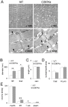
Figure 5. The SR in skeletal muscles of CD97Ko mice is morphologically abnormal.
A Representative electron micrographs of longitudinal ultrathin sections of WT and CD97Ko mice. Note the altered morphology of the SR which was frequently swollen (arrow heads), whereas myofibrils and mitochondria retained normal structures in CD97Ko mice. B-E Quantitative measurements of several morphometric parameters based on electron micrographs of the quadriceps muscle (n = 4 mice/strain, 25 micrographs/animal, mean ± SEM; *p<0.05, **p<0.01, *** p<0.001, t-test). D shows the number of AI-junctions and triads/100 µm2 area occupied by myofibrils.
Figure 6
Figure 6. Macroscopic and microscopic features of adult CD97Ko and WT skeletal muscles are comparable.
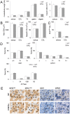
A-E 10 mice per strain were analyzed; data are mean ± SEM (t-test). A Percentage of muscle to body weight and cross-sectional area (CSA) of several skeletal muscles: extensor digitorum longus (EDL), tibialis anterior (TA), gastrocnemius (gastro), digastricus (digastr). B Cross-sectional area (CSA) of single fibers (n = 30/mouse) and number of fibers/area (n = 2 areas/muscle) of soleus and EDL muscles. C Percentage of MHCslow-positive fibers within the EDL and soleus muscles stained by immunohistology. D Histochemistry for succinate dehydrogenase (SDH), glycerol-3-phosphate-dehydrogenase (GPDH), and ATPase (pH 4.5) in soleus muscles. Staining intensities were quantified according to visible subtypes. E Physiologic and metabolic profile of murine soleus muscles of a WT and CD97Ko mouse; 20 fibers were identified and typed in each section. WT mouse (fibers 1-3, 6-8, 10, 14, and 18 are MHCslow-positive, all fibers have the metabolic phenotype slow-oxidative; fibers 4, 5, 9, 12, 13, 15–17, 19, and 20 are MHCfast-positive, metabolic phenotype fast-oxidative glycolytic II; fiber 11 is MHCfast/slow-double-positive, metabolic phenotype slow-oxidative); CD97 Ko mouse (fibers 7, 10, 11, 14, and 17–20 are MHCslow-positive, metabolic phenotype slow-oxidative; fibers 1, 3–6, 8, 9, 12, 13, 15, and 16 are MHCfast-positive, metabolic phenotype fast-oxidative glycolytic II; fiber 2 is MHCfast/slow-double-positive, metabolic phenotype slow-oxidative).
Figure 7
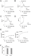
Figure 7. Calcium release from myofibers and muscle force generation and fatigability are normal in CD97Ko mice.
A–D Intracellular Ca2+ release from the SR into cytoplasm of single muscle flexor digitorum brevis (FDB) myofibers. Mean traces of the analyzed FDB fibers from adult WT and CD97Ko represent [Ca2+]i responses measured by Fura-2AM video microscopy. Variations in [Ca2+]i over time are represented by the ratio fluorescence intensities (FI) at 340 and 380 nm excitation wavelengths after dynamic background subtraction. Fibers were exposed to calcium release activators after 60 s. From 120 s till 300 s, medium without a substance was applied again. 5 mice per strain and from each mouse two to three fibers were analyzed (mean ± SEM, Mann-Whitney test). A Basal 340/380 fluorescence intensities (FI) ratios were unchanged in both WT and CD97ko fibers. B, C 100 mM KCl (B) and 30 mM caffeine (C) increased [Ca2+]i comparably in WT and CD97Ko fibers. D Application of 5 µM ionomycin as a positive control induced a fast total store [Ca2+]i release from the SR. E, F Functional analysis of skeletal muscles of CD97Ko and WT mice at the age of 2 and 4 months. Force-frequency relationship (E) and muscle fatigability, determined as percentage of decline in force over 15 s (F), were measured in soleus muscles (n = 8 mice/strain; mean ± SEM, t-test).
Figure 8
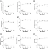
Figure 8. Unchanged wheel-running activity of CD97Ko mice.
Measures of wheel-running activity of CD97Ko and WT mice (n = 16 mice/strain; mean ± SEM; t-test, *p<0.05). Running for 24 h was repeated every 2 weeks. A–E Average and maximum acceleration (A), duration/run and over 24 h (B), average and maximum speed (C), and distance/run or over 24 h (D) were measured. Body weight was monitored (E).

Similar articles

Cited by

References

    1. Yona S, Lin HH, Siu WO, Gordon S, Stacey M (2008) Adhesion-GPCRs: emerging roles for novel receptors. Trends Biochem Sci 33: 491–500. - PubMed
    1. Langenhan T, Aust G, Hamann J (2013) Sticky signaling - Adhesion class G protein-coupled receptors take the stage. Sci Signal 6: re3 doi: 10.1126/scisignal.2003825 - DOI - PubMed
    1. Lin HH, Chang GW, Davies JQ, Stacey M, Harris J, et al. (2004) Autocatalytic cleavage of the EMR2 receptor occurs at a conserved G protein-coupled receptor proteolytic site motif. J Biol Chem 279: 31823–31832. - PubMed
    1. Arac D, Boucard AA, Bolliger MF, Nguyen J, Soltis SM, et al. (2012) A novel evolutionarily conserved domain of cell-adhesion GPCRs mediates autoproteolysis. EMBO J 31: 1364–1378. - PMC - PubMed
    1. Eichler W, Aust G, Hamann D (1994) Characterization of the early activation-dependent antigen on lymphocytes defined by the monoclonal antibody BL-Ac(F2). Scand J Immunol 39: 111–115. - PubMed

Publication types

MeSH terms