. 2014 Jun 19;157(7):1605-18.
doi: 10.1016/j.cell.2014.04.033.

Programmed translational readthrough generates antiangiogenic VEGF-Ax

Affiliations

Programmed translational readthrough generates antiangiogenic VEGF-Ax

Sandeepa M Eswarappa et al. Cell. .

Abstract

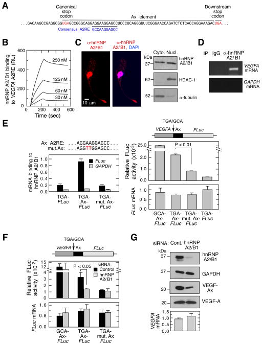
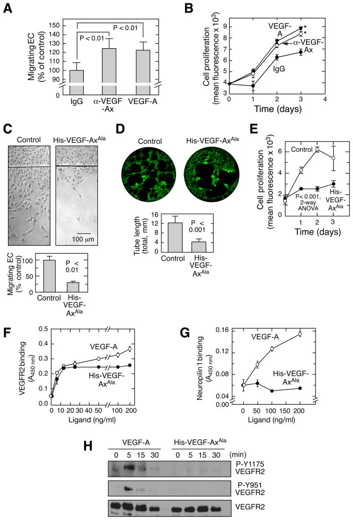
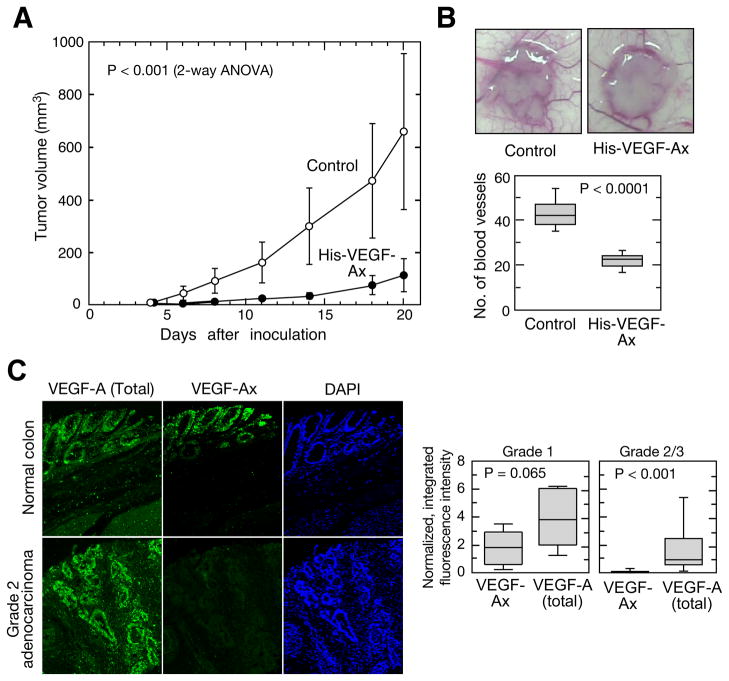
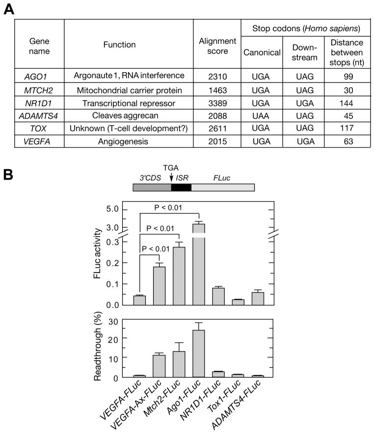
Translational readthrough, observed primarily in less complex organisms from viruses to Drosophila, expands the proteome by translating select transcripts beyond the canonical stop codon. Here, we show that vascular endothelial growth factor A (VEGFA) mRNA in mammalian endothelial cells undergoes programmed translational readthrough (PTR) generating VEGF-Ax, an isoform containing a unique 22-amino-acid C terminus extension. A cis-acting element in the VEGFA 3' UTR serves a dual function, not only encoding the appended peptide but also directing the PTR by decoding the UGA stop codon as serine. Heterogeneous nuclear ribonucleoprotein (hnRNP) A2/B1 binds this element and promotes readthrough. Remarkably, VEGF-Ax exhibits antiangiogenic activity in contrast to the proangiogenic activity of VEGF-A. Pathophysiological significance of VEGF-Ax is indicated by robust expression in multiple human tissues but depletion in colon adenocarcinoma. Furthermore, genome-wide analysis revealed AGO1 and MTCH2 as authentic readthrough targets. Overall, our studies reveal a novel protein-regulated PTR event in a vertebrate system.


Conflict of interest statement

None of the other authors have any financial conflict of interest with the information in this manuscript.

Figures

Figure 1
Figure 1. ECs Express and Secrete an Anti-angiogenic Isoform of VEGF-A
(A) Neutralizing anti-VEGF-A antibody enhances EC migration. Shown are ECs migrating across razor-wound line (mean ± SE, n=3) (left), individual cell trajectories and root-mean-square displacement (center), and cell speed in the presence of anti-VEGF-A antibody (n=71) and IgG (n=52) (right). (B) Neutralizing anti-VEGF-A antibody increases EC proliferation. Cell amount was determined fluorimetrically as DNA (mean ± SE, n=4). (C) EC-derived conditioned medium inhibits EC migration. EC migration determined by razor-wound assay (mean ± SE, n=3). (D) Conservation of VEGFA 3′UTR proximal region (gray); canonical and downstream, in-frame stop codons in mammals (boxed). (E) Schematic showing generation of VEGF-A, VEGF-Ab, and VEGF-Ax isoforms. See also Figure S1.
Figure 2
Figure 2. VEGF-Ax is a Novel Isoform of VEGFA Generated by Translational Readthrough
(A) VEGFA-specific siRNAs inhibit VEGF-Ax expression. Bovine ECs were transfected with three different VEGFA-specific siRNAs and VEGF-Ax in cell lysates determined by immunoblot with anti-VEGF-Ax antibody. (B) VEGF-Ax is an authentic VEGF-A isoform. Lysates from bovine EC were immunoprecipitated with anti-VEGF-Ax antibody or pre-immune (Pre-imm.) serum and subjected to immunoblot analysis with anti-VEGF-Ax and anti-VEGF-A antibodies. (C) VEGF-Ax is expressed by murine aortic ECs (MAEC), bovine aortic ECs (BAEC), and human umbilical vein ECs (HUVEC). Cell lysates were subjected to immunoblot analysis with anti-VEGF-Ax antibody. (D) Separation of VEGF-Ax and VEGF-A isoforms in EC. BAEC lysates (left) and conditioned media (right) were deglycosylated and resolved on 16% Tricine gel before immunoblot analysis with anti-VEGF-Ax and anti-VEGF-A antibodies. (E) Amino acid sequence of VEGF-Ax. Peptides identified by mass spectrometry (underline), readthrough region (bold), and recoded Ser (boxed) are highlighted. (F) Identification of VEGFA mRNA readthrough product by MS/MS. A MS/MS spectrum was identified consistent with RSAGLEEGASLR (readthrough amino acid is underlined). The spectrum contains a total of nine C-terminal ‘y’ ions and seven N-terminal ‘b’ ions consistent with this sequence. The peptide was a low abundant component, and the spectrum contains several contaminant ions (*). (G) Identity of readthrough peptide validated by synthetic peptide. RSAGLEEGASLR peptide was synthesized and the MS/MS spectrum determined as in (F). See also Figure S2.
Figure 3
Figure 3. Ax Element is Necessary and Sufficient cis-acting Signal for VEGFA mRNA Readthrough
(A) Ax element is sufficient for readthrough of VEGFA chimeric transcript. Plasmid containing in-frame VEGF-Ax-FLuc, and variants with TGA-to-GCA substitution, no Ax element, and Ax replaced by a non-specific sequence (ns), were transfected into ECs. FLuc activity was normalized to expression of co-transfected Renilla Luc (top), and FLuc mRNA expression determined by qRT-PCR (bottom). (B) Ax element is sufficient for readthrough of heterologous mRNA, Chimeric plasmids containing Myc-Ax-FLuc and variants were transfected into ECs. Relative FLuc activity and mRNA expression were measured (left). Readthrough product also was determined by immunoprecipitation with rabbit anti-Myc-tag antibody followed by immunoblot with the mouse anti-Myc-tag antibody (right). (C) Nucleotide sequence of bovine Ax element. Flanking stop codons (boxes), deletions (horizontal lines) and mutations (arrows) are shown. (D) Plasmids containing deletions of VEGF-Ax-FLuc were transfected into ECs. FLuc activity was normalized by expression of co-transfected Renilla Luc (top); FLuc mRNA expression was determined by qRT-PCR (bottom). (E) All three stop codons permit readthrough. ECs were transfected with VEGF-Ax-FLuc constructs containing each stop codon. Relative FLuc activity and mRNA were determined. (F) The nucleotide immediately following the canonical stop codon (G1) does not alter readthrough efficiency. ECs were transfected with VEGF-Ax-FLuc constructs containing G-to-C, G-to-A, and G-to-T substitutions. Relative FLuc activity and mRNA were measured. See also Figure S3.
Figure 4
Figure 4. hnRNP A2/B1 Facilitates VEGFA mRNA Translational Readthrough
(A) Inter-stop codon sequence (Ax element) of human VEGFA contains a near-consensus hnRNP A2/B1 response element (A2RE). (B) High-affinity binding of hnRNP A2/B1 to VEGFA A2RE. Surface plasmon resonance analysis of recombinant hnRNP A2/B1 binding to biotinylated RNA containing VEGFA A2RE immobilized on streptavidin sensor chip (in response units, RU). (C) Cytoplasmic localization of hnRNP A2/B1 in ECs. Detection of hnRNP A2/B1 by immunofluorescence (left, with nuclear DAPI stain) and by immunoblot of fractionated compartments (right). (D) hnRNP A2/B1 binds VEGFA mRNA in cells. Immunoprecipitation of hnRNP A2/B1 followed by RT-PCR using VEGFA- and GAPDH-specific primers. (E) hnRNP A2/B1 binds the A2RE in VEGFA mRNA. VEGF-Ax-FLuc plasmid containing an Ax element with A2RE mutation (mut. Ax) was transfected into ECs. Anti-hnRNP A2/B1 immunoprecipitates were probed by qRT-PCR using FLuc- and GAPDH-specific TaqMan probes (left). Readthrough expressed as relative FLuc activity and mRNA level are shown (right). (F) siRNA-mediated knockdown of hnRNP A2/B1 reduces readthrough of Ax element-containing plasmid. ECs were transfected with VEGF-Ax-FLuc construct and the variant containing A2RE mutation. FLuc activity was measured and FLuc mRNA determined by qRT-PCR. (G) siRNA-mediated knockdown of hnRNP A2/B1 reduces VEGF-Ax expression. Following siRNA-mediated knockdown of hnRNP A2/B1, VEGF-Ax and VEGF-A were determined by immunoblot analysis and VEGFA mRNA by qRT-PCR.
Figure 5
Figure 5. Anti-angiogenic properties of VEGF-Ax
(A,B) Anti-VEGF-Ax antibody inhibits EC migration and proliferation. EC migration (A) in the presence of anti-VEGF-Ax antibody or recombinant human VEGF-A (20 ng/ml) was measured by razor-wound assay. Cell proliferation (B) was measured fluorimetrically as DNA (*, P < 0.05, 2-way ANOVA). (C–E) Anti-angiogenic properties of VEGF-Ax. Migration (C), tube formation in Matrigel (D), and proliferation (E) of bovine ECs in the presence of recombinant His-VEGF-Axala (50 ng/ml). (F–G) Binding of VEGF-A and His-VEGF-Axala to VEGFR2 (F) and neuropilin-1 (G). Binding was measured colorimetrically by solid phase enzyme-linked receptor-binding assay. (H) Phosphorylation status of VEGFR2. HUVECs were treated with VEGF-A and His-VEGF-AxAla for up to 30 min and immunoblotted as shown. See also Figure S4.
Figure 6
Figure 6. Anti-tumor properties of VEGF-Ax
(A) Tumor progression in athymic nude mice. Mice were inoculated with HCT116 cells and treated sub-cutaneously with recombinant His-VEGF-Ax (10 μg/mouse; n=6 mice, 12 tumors) or buffer (n=5 mice, 10 tumors) every third day starting from 4th day after tumor cell inoculation. (B) Tumor angiogenesis. Representative images of day 6 HCT116 tumors in athymic nude mice (top). Number of blood vessels feeding directly into tumors; whiskers show 5th and 95th percentiles (bottom; n=6 mice, 12 tumors in each group). (C) Immunofluorescence of normal colon and colon adenocarcinoma. VEGF-Ax and total VEGF-A were quantified as integrated, background-subtracted fluorescence intensity (left); expression in Grade 1 (n=6) and Grade 2/3 colon adenocarcinoma (n=22) were normalized to healthy colon (n=5); whiskers show 5th and 95th percentiles (right). See also Figure S5.
Figure 7
Figure 7. Genome-wide Analysis to Identify Targets of Translational Readthrough
(A) Properties of candidate genes selected for experimental validation. Candidates were selected using a bioinformatic screening protocol (Figure S6). (B) Validation of translational readthrough targets. Chimeric plasmids containing about 700 nt of the 3′-terminus of human coding sequences upstream of the inter-stop codon region and in-frame with FLuc were transfected into HEK293 cells. Relative FLuc activities normalized to FLuc mRNA were determined by qRT-PCR (top). Readthrough efficiency is expressed as percent of FLuc activity of normal translation controls in which canonical UGA stop codon is replaced by GCA (bottom). See also Figure S6.

Comment in

  • Cancer genetics: Ignoring the signs.
    Lokody I. Lokody I. Nat Rev Cancer. 2014 Aug;14(8):514. doi: 10.1038/nrc3787. Epub 2014 Jul 10. Nat Rev Cancer. 2014. PMID: 25008265 No abstract available.

References

    1. Ausprunk DH, Folkman J. Migration and proliferation of endothelial cells in preformed and newly formed blood vessels during tumor angiogenesis. Microvasc Res. 1977;14:53–65. - PubMed
    1. Bates DO, Cui TG, Doughty JM, Winkler M, Sugiono M, Shields JD, Peat D, Gillatt D, Harper SJ. VEGF165b, an inhibitory splice variant of vascular endothelial growth factor, is down-regulated in renal cell carcinoma. Cancer Res. 2002;62:4123–4131. - PubMed
    1. Bates DO, Mavrou A, Qiu Y, Carter JG, Hamdollah-Zadeh M, Barratt S, Gammons MV, Millar AB, Salmon AH, Oltean S, et al. Detection of VEGF-A(xxx)b isoforms in human tissues. PloS ONE. 2013;8:e68399. - PMC - PubMed
    1. Beier H, Grimm M. Misreading of termination codons in eukaryotes by natural nonsense suppressor tRNAs. Nucleic Acids Res. 2001;29:4767–4782. - PMC - PubMed
    1. Carmeliet P, Ferreira V, Breier G, Pollefeyt S, Kieckens L, Gertsenstein M, Fahrig M, Vandenhoeck A, Harpal K, Eberhardt C, et al. Abnormal blood vessel development and lethality in embryos lacking a single VEGF allele. Nature. 1996;380:435–439. - PubMed

Publication types

Substances