. 2014 Jun 19;157(7):1657-70.
doi: 10.1016/j.cell.2014.04.035.

Conservation of Ca2+/calmodulin regulation across Na and Ca2+ channels

Affiliations

Conservation of Ca2+/calmodulin regulation across Na and Ca2+ channels

Manu Ben-Johny et al. Cell. .

Abstract

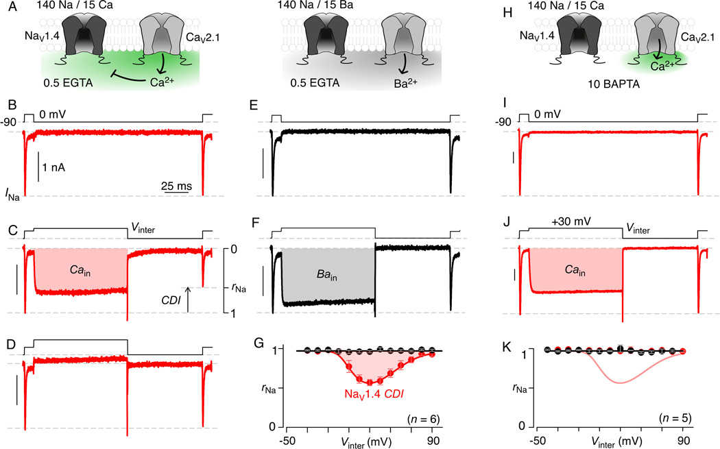
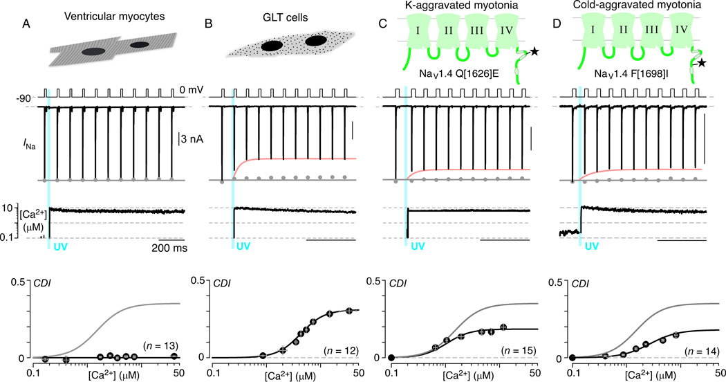
Voltage-gated Na and Ca2+ channels comprise distinct ion channel superfamilies, yet the carboxy tails of these channels exhibit high homology, hinting at a long-shared and purposeful module. For different Ca2+ channels, carboxyl-tail interactions with calmodulin do elaborate robust and similar forms of Ca2+ regulation. However, Na channels have only shown subtler Ca2+ modulation that differs among reports, challenging attempts at unified understanding. Here, by rapid Ca2+ photorelease onto Na channels, we reset this view of Na channel regulation. For cardiac-muscle channels (NaV1.5), reported effects from which most mechanistic proposals derive, we observe no Ca2+ modulation. Conversely, for skeletal-muscle channels (NaV1.4), we uncover fast Ca2+ regulation eerily similar to that of Ca2+ channels. Channelopathic myotonia mutations halve NaV1.4 Ca2+ regulation, and transplanting the NaV1.4 carboxy tail onto Ca2+ channels recapitulates Ca2+ regulation. Thus, we argue for the persistence and physiological relevance of an ancient Ca2+ regulatory module across Na and Ca2+ channels.


Figures

Figure 1
Figure 1. Homology but divergent function for Ca2+versus Na channels
(A) CI region of Ca2+ (CaV1.3) and Na channels (NaV1.5, NaV1.4). Dual vestigial EF-hands shaded in rose and green. IQ domain, blue. (B) Ca2+ channel regulation inducible by channel Ca2+ influx. Ba2+ influx as negative control. (C) Left, CaV1.3 current traces carried by Ca2+(red) or Ba2+(black). Vertical bar for Ca2+ trace. Ba2+ trace scaled ~3× downward for kinetic comparison. Right, r300 (fraction of peak current remaining after 300-ms depolarization) versus Vtest potential, plotted as mean ± SEM (8 cells). (D) Na channels characterized under pipet dialysis with 0 or 10 µM Ca2+. (E) Schematic of reported Ca2+ effects on inactivation. Left, h, fractional current remaining after prepulses (Vhold). Right, purported Ca2+-induced voltage shifts of h. (F) NaV1.5 currents under protocol in panel E (black, 0 Ca2+; red, 10 µMCa2+buffered with HEDTA). See Figure S1. (G, H) Normalized form of h unaffected by Ca2+. Potential Ca2+-induced reduction in NaV1.4 h (rose dashed line). Error bars, SEM throughout. Fit function: h = 1 / (1 + exp ((VholdV1/2) / SF), where SF = 6.2 (NaV1.4) and 7.5 (NaV1.5).
Figure 2
Figure 2. CDI of Na channels under Ca2+photouncaging
(A) NaV1.5 currents unaffected by 10 µM Ca2+. Gray dots, peak currents before uncaging. Bottom, mean data for CDI versus Ca2+-step amplitude.CDI = 1 – average peak INa of last 3–4 responses after Ca2+ uncaging / peak INa before uncaging. Symbols, mean ± SEM of ~5 uncaging events compiled from 23 cells. See Figure S2. (B) NaV1.4 peak currents decline during 2 µM Ca2+ step (rose fit). Format as in panel A. Bottom, mean CDI plotted versus Ca2+. Each symbol, mean ± SEM of ~5 uncaging events compiled from 35 cells. See Figures S2–S4. (C) NaV1.4 currents specifying h at ~100 nM Ca2+. Bottom, h curve (mean ± SEM, 5 cells). (D)~3 µM Ca2+ step uniformly suppresses Na currents. Bottom, corresponding mean h curve (red symbols and fit), where symbols plot mean ± SEM (5 cells). Upwardly scaled h curve (rose) same as before uncaging (black).
Figure 3
Figure 3. Na channel regulated by Ca2+spill over from Ca2+ channels
(A) Schematic, Ca2+ spill over from CaV2.1 inhibiting Na channels. (B) Dual voltage-pulses selectively evoke identical NaV1.4 currents. See Figure S5. (C) Intervening +30 mV pulse (Vinter) activates CaV2.1, diminishing ensuing Na current. rNa, fraction of Na current remaining after CaV2.1 Ca2+ influx. (D) Vinter to +90 mV rescues the second NaV1.4 current. (E, F) Na current amplitude unperturbed by Ba2+ influx through CaV2.1 channels. (G) Mean relation for rNa versus Vinter shows U-shape with Ca2+ (red) but not Ba2+ (black). Symbols, mean ± SEM (6 cells). (H–K) Restricting Ca2+ to CaV2.1nanodomain prevents Na channel CDI. Panel K, symbols, mean ± SEM (5 cells), format as in panel G. See Figure S5.
Figure 4
Figure 4. Multi-channel stochastic records of NaV1.4 CDI
(A) Multi-channel records from HEK293 cells coexpressing NaV1.4 and CaV2.1 channels. On-cell patch configuration. Voltage protocol (top), multi-channel record (middle), and ensemble average current (bottom). Red shading, Ca2+ entry. Ensemble average shows reduced Na channel activity after Ca2+ entry (pulse ii) versus before (pulse i). (B) Amplitude histogram analysis of patch from panel A shows no change in unitary current following Ca2+ entry (top before interpulse; bottom after interpulse). Amplitude histogram analysis of events occuring 0.5–17 m sec after pulse onset during −30 mV test pulses. Fits (black) to data (gray) derived from amplitude analysis of low-pass filtered stochastic channel simulations with added gaussian noise. Dashed-red lines, unitary current i used to generate fits. (C) Expanded time base display of ensemble average currents from panel A, before (upper) and after (below) Ca2+. Fast inactivation essentially identical; same time constant for both mono exponential fits (black curves). (D) Multi-channel stochastic records of separate patch with only NaV1.4 channels. No difference in channel activity before and after intervening pulse (mean decrement ~ 0.2 ± 3% (mean ± SEM, n = 5 patches). Second multi-channel record chosen to illustrate rare occurrence of persistent gating mode. Format as in panel A.
Figure 5
Figure 5. Calmodulin as Ca2+ sensor for NaV1.4CDI
(A) Mutating putative Ca2+-coordinating residues in NaV1.4 EF-hand did not alter CDI. Format as in Figure 2A. Symbols, mean ± SEM of ~3 uncaging events from 12 cells. (B) CaM1234 abolishes CDI. Symbols, mean ± SEM (~6 uncaging events from 27 cells). (C) Eliminating N-lobe Ca2+-binding (CaM12) abolishes CDI. Symbols, mean ± SEM of 4–5 uncaging events from 12 cells. (D) Eliminating C-lobe Ca2+ binding (CaM34) spares CDI. Symbols, mean ± SEM of ~5 uncaging events from 12 cells. (E) Mutating NaV1.4 III–IV loop spares CDI. Format as in Figure 2A. Symbols, mean ± SEM of 4–5 uncaging events from 18 cells. (F) No CDI in NaV1.4–1.5CT chimera. Symbols, mean ± SEM of 4–5 uncaging events (13 cells). (G) Substituting dual alanines for key isoleucine-glutamine residues in NaV1.4 IQ domain yields facilitating Na currents. Bottom, mean data confirms facilitation, shown as negative CDI. Symbols, mean ± SEM of ~13 uncaging events (20 cells).
Figure 6
Figure 6. Physiology of Na channel Ca2+ regulation
(A) No Ca2+-regulation of native NaV1.5 in ventricular myocytes. Format as in Figure 2A. Symbols, mean ± SEM from 5–6 uncaging events (13 cells). (B) Endogenous NaV1.4 channels in GLT cells exhibit CDI. Minimal contamination by Ca2+-activated Cl current (<5% of INa) subtracted. Each symbol, mean ± SEM from 4–5 uncaging events (12 cells). (C, D) Recombinant NaV1.4 channels with mutations for K- and cold-aggravated myotonias show weaker CDI. Symbols, mean ± SEM from ~9 uncaging events (indicated number of cells).
Figure 7
Figure 7. Persistence of CaM/CI module across Na and Ca2+channel superfamilies
(A) Transferring NaV1.4 carboxy tail to NaV1.5 backbone (NaV1.5–1.4CT) confers latent Ca2+regulation (wild-type NaV1.4, gray fit in bottom subpanel). Format as in Figure 2A. Symbols, mean ± SEM from 4–5 uncaging events (10 cells). (B) Phylogenetic tree of the Na and Ca2+ channel superfamilies. (C) Transplanting NaV1.4 carboxy tail onto CaV1.3 backbone (CaV1.3–NaV1.4CT) yields chimeric channel that retains Ca2+ regulation. Format as in Figure 1C. Symbols, mean ± SEM, 7 cells. CDI measured under low Ca2+ buffering (see Extended Experimental Procedures). (D) Coexpressimg CaM1234with CaV1.3–NaV1.4 CT abolishes CDI. Format as in Figure 1C. Symbols, mean ± SEM, 7 cells. CDI measured as in panel C. (E) Maximum likelihood phylogenetic tree shows conservation among Ca2+ and Na channel CI regions, across major eukaryotic phyla. Format as in Figure 1A. Consensus sequence patterns for motifs on top. Sequence alignment starts at the center with the Paramecium Ca2+ channel. Ca2+ channels from progressively more advanced organisms branch to the top (pale colors), and those for Na channels branch to the bottom (darker colors).

References

    1. Adams PJ, Snutch TP. Calcium channelopathies: voltage-gated calcium channels. Sub-cellular biochemistry. 2007;45:215–251. - PubMed
    1. Alseikhan BA, DeMaria CD, Colecraft HM, Yue DT. Engineered calmodulins reveal the unexpected eminence of Ca2+ channel inactivation in controlling heart excitation. Proc Natl Acad Sci U S A. 2002;99:17185–17190. - PMC - PubMed
    1. Babitch J. Channel hands. Nature. 1990;346:321–322. - PubMed
    1. Bazzazi H, Ben Johny M, Adams PJ, Soong TW, Yue DT. Continuously Tunable Ca(2+) Regulation of RNA-Edited CaV1.3 Channels. Cell Rep. 2013;5:367–377. - PMC - PubMed
    1. Ben Johny M, Yang PS, Bazzazi H, Yue DT. Dynamic switching of calmodulin interactions underlies Ca2+ regulation of CaV1.3 channels. Nature communications. 2013;4:1717. - PMC - PubMed

Publication types

LinkOut - more resources