. 2014 Jun 19;157(7):1671-84.
doi: 10.1016/j.cell.2014.04.037.

Structure of a RING E3 trapped in action reveals ligation mechanism for the ubiquitin-like protein NEDD8

Affiliations

Structure of a RING E3 trapped in action reveals ligation mechanism for the ubiquitin-like protein NEDD8

Daniel C Scott et al. Cell. .

Abstract

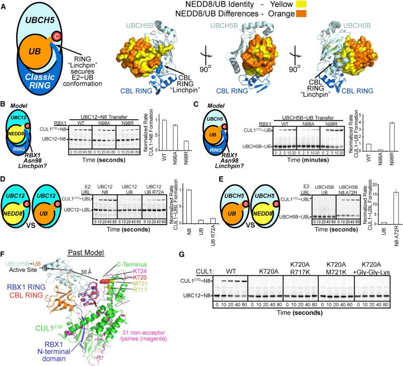
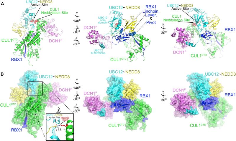
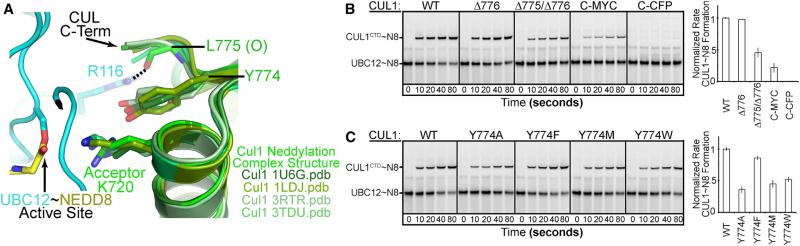
Most E3 ligases use a RING domain to activate a thioester-linked E2∼ubiquitin-like protein (UBL) intermediate and promote UBL transfer to a remotely bound target protein. Nonetheless, RING E3 mechanisms matching a specific UBL and acceptor lysine remain elusive, including for RBX1, which mediates NEDD8 ligation to cullins and >10% of all ubiquitination. We report the structure of a trapped RING E3-E2∼UBL-target intermediate representing RBX1-UBC12∼NEDD8-CUL1-DCN1, which reveals the mechanism of NEDD8 ligation and how a particular UBL and acceptor lysine are matched by a multifunctional RING E3. Numerous mechanisms specify cullin neddylation while preventing noncognate ubiquitin ligation. Notably, E2-E3-target and RING-E2∼UBL modules are not optimized to function independently, but instead require integration by the UBL and target for maximal reactivity. The UBL and target regulate the catalytic machinery by positioning the RING-E2∼UBL catalytic center, licensing the acceptor lysine, and influencing E2 reactivity, thereby driving their specific coupling by a multifunctional RING E3.


Figures

Figure 1
Figure 1. Specificity of RBX1-Mediated NEDD8 Ligation to CUL1
(A) Canonical RING-E2~UB architecture, highlighting the linchpin and conservation of E2 and RING binding residues in UB and NEDD8, shown for CBL-UBCH5~UB (Dou et al., 2013). (B) Mutational analysis of RBX1's Asn98, corresponding to canonical RING linchpin, in RBX1-mediated pulse-chase fluorescent NEDD8 transfer from UBC12 to CUL1CTD. Graph, rate compared to wild-type (WT) RBX1; error, 1 SD. (C) Mutational analysis of RBX1's Asn98, aka canonical RING linchpin, in RBX1-mediated pulse-chase fluorescent UB transfer from UBCH5B to CUL1CTD. Graph, rate compared to WT RBX1; error, 1 SD. (D) Role of UBL in RBX1-mediated CUL1 modification assayed by comparing pulse-chase NEDD8, UB, or UB R72A transfer from NEDD8's E2 UBC12. The noncognate UBL UB was loaded on UBC12 in the pulse reaction either through its R72A mutation or use of E1 mutant UBA3 R190Q. Graph, rate compared to WT UBC12~NEDD8; error, 1 SD. (E) Role of UBL in RBX1-mediated CUL1 modification assayed by comparing UB or NEDD8 A72R transfer from UBCH5B to CUL1CTD. The NEDD8 A72R mutation allows loading this noncognate UBL on UBCH5B in the pulse reaction. Graph, rate compared to WT UBCH5~UB; error, 1 SD. (F) CUL1CTD Lys locations. Docking RBX1's RING from prior structures with that from CBL-UBCH5~UB revealed a >30Å gap between the active site and K720 (red) or other lysines (native in magenta, introduced in yellow) in CUL1 (Dou et al., 2013; Zheng et al., 2002). (G) Acceptor Lys specificity for NEDD8 transfer from UBC12 to CUL1CTD or Lys mutants. Gly-Gly-Lys, appended to C terminus of CUL1CTD K720A.
Figure 2
Figure 2. Structure of RBX1-UBC12~NEDD8-CUL1CTD-DCN1P —A RING E3 in Action
(A) Structure of neddylation complex as cartoon, with active site and CUL1 acceptor residue 720 in red, and RBX1 “linchpin, lever, and pivot” spheres in center panel. (B) Surface representation. Inset: model of catalytic center. See also Figure S1 and Movie S1.
Figure 3
Figure 3. RING-E2 UBL-Acceptor Modules for Neddylation
(A) Different RING-E2~UBL closed conformations shown with the E2 catalytic core domain aligned for RBX1-UBC12~NEDD8-CUL1CTD-DCN1P and CBL-UBCH5~UB (Dou et al., 2013). (B) Close-up of E2~UBL interfaces from (A). (C) Variation in E2~UBL interfaces shown with UBLs aligned from RBX1-UBC12~NEDD8-CUL1CTD-DCN1P and CBL-UBCH5~UB. (D) Pulse-chase assays for compensation between UBC12 Leu mutants and NEDD8 I44A in RBX1-mediated CUL1 modification. (E) Pulse-chase assays testing compensation between UBCH5 Leu mutant and UB I44A in RBX1-mediated CUL1 modification. (F) Different RING-E2~UBL active site presentations highlighted by relative placement of UBC12 D143 and UBCH5B D117 side chains, viewed with UBLs aligned from RBX1-UBC12~NEDD8-CUL1CTD-DCN1P (CUL1 acceptor K720 modeled in place of Arg) and CBL-UBCH5~UB. UBC12 D143 carbonyl is also shown in sticks. (G) Different E2~UBL active site presentations in UBC12~NEDD8-CUL1 and UBC9-SUMO~RANGAP1 (Reverter and Lima, 2005), oriented by aligning UBC9 on UBCH5 from (F) and highlighting different relative positions of UBC12 D143 and UBC9 D127. UBC12 D143 carbonyl is also shown in sticks. (H) RBX1-mediated pulse-chase fluorescent NEDD8 transfer from UBC12 to CUL1CTD testing roles of side chains from RBX1's N113, which aligns NEDD8's C-terminal tail, and D143, opposite the UBC12~NEDD8 bond. (I) RBX1's distinct linchpin Arg46 in spheres, shifted across RING domain from location of canonical linchpin as typified in CBL. RING-E2~UBL portions of RBX1-UBC12~NEDD8-CUL1CTD-DCN1P and CBL-UBCH5~UB structures shown with UBLs aligned. (J) Linchpins (blue) in human RING domain sequences. Red, zinc ligands; green, E2-binding residue; purple, RBX1 pivot; yellow, lever. (K) RBX1-mediated pulse-chase fluorescent NEDD8 transfer from UBC12 to CUL1CTD testing role of RBX1 R46 linchpin. (L) Pulse-chase assays testing role of NEDD8's Leu8 and Thr9 from the b1-b2 loop in RBX1-mediated transfer from UBC12 to CUL1CTD. See also Figure S2.
Figure 4
Figure 4. NEDD8 Pushes an RBX1 Lever and Directs an RBX1 Pivot to Juxtapose the Active Site and Acceptor Lys
(A) RBX1 RING domain and W35 “pivot” positions in neddylation complex compared with CUL1-RBX1-CAND1 (Goldenberg et al., 2004; Zheng et al., 2002), with CUL1 aligned. (B) Close-up of contacts between UBC12-linked donor NEDD8 and RBX1 linker. Zinc atoms bound to RING are shown as spheres. (C) As in (B) except with NEDD8 in surface colored by identity with UB. RING and UBC12-binding residues are identical between NEDD8 and UB (yellow), but exposed surfaces and contacts to RBX1 pivot differ (orange). (D) Comparison of effects of Ala mutations in place of RBX1 “lever” (I37) or other linker residues in RBX1-mediated NEDD8 transfer from UBC12 to CUL1CTD. Graph, rate compared to WT RBX1; error, 1 SD. (E) Same as (D) but with variants of RBX1 Trp35 pivot. (F) Same as (E) but monitoring fluorescent UB transfer from UBCH5B. (G) Immunoblot comparing RBX1-mediated transfer of WT NEDD8, or variants with corresponding E31Q and E32D from UB, from UBC12 to CUL1CTD. Graph, rate compared to WT UBC12~NEDD8; error, 1 SD. (H) RBX1-mediated transfer of fluorescent UB, or “neddylized” variants with Q31E and D32E, from UBCH5B to CUL1CTD. Graph, rate compared to WT UBCH5~UB; error, 1 SD. (I) Schematic of roles and orientations of RBX1 elements and interactions with UBC12Pulse-chase assays forNEDD8, oriented as in prior RBX1-CUL1 structures or RBX1-UBC12~NEDD8-CUL1CTD-DCN1P. (J and K) Pulse-chase assays for NEDD8-modifed SCFFbw7ΔD testing roles of RBX1 W35 “pivot” (J) and I37 “lever” (K) in directing WT or “neddylized” Q31E/D32E UB from UBCH5B to either an F-box-bound substrate (Cyclin E phosphopeptide) or neddylated CUL1. See also Figure S3 and Movie S2.
Figure 5
Figure 5. CUL1 Features Contributing to NEDD8 Acceptor Lys Selection
(A) Close-up of complementary UBC12-CUL1 interface. Note UBC12 proximity to CUL1's C terminus and interactions with carbonyl from CUL1's penultimate Leu775. Tyr774 structurally aligns CUL1's Lys720, even in the absence of neddylation enzymes as in superposition of prior CUL1 WHB domain structures. (B) Role CUL1 C terminus forming a complementary surface with UBC12, assayed by pulse-chase RBX1-mediated transfer of fluorescent NEDD8 from UBC12 to the indicated CUL1CTD C-terminal deletion or extension variants. Graph, rate compared to CUL1; error, 1 SD. (C) Effects of substituting the CUL1 Lys720 stabilizing residue Tyr774 in experiments as in (B).
Figure 6
Figure 6. DCN1P Synergizes with Catalytic RBX1-UBC12~NEDD8-CUL1 Architecture to Promote Neddylation
(A) RBX1-UBC12~NEDD8-CUL1CTD-DCN1P structure, highlighting complementarity of NEDD8's surface with structure of RBX1 and UBC12 core domain, and unstructured UBC12 region linking UBC12's catalytic E2 core domain with the acetylated N terminus bound to DCN1P. (B) Incompatibility of DCN1-UBC12~NEDD8 binding to RBX1-CUL1-CAND1 due to NEDD8 clashing with RBX1 and CUL1. Model generated by docking RING domains from the present and prior 1U6G Protein Data Bank (PDB) structures, with RBX1NTD-CUL1CTD oriented as in (A). (C) Incompatibility of DCN1-UBC12~NEDD8 binding to RBX1-CUL1 in conformation from 3RTR PDB due to NEDD8 clashing with RBX1-CUL1, and DCN1-UBC12 clashing with RBX1. Model generated by docking RING domains from the present and prior 3RTR PDB structures, with RBX1NTD-CUL1CTD oriented as in (A). (D) Incompatibility of DCN1-UBC12~NEDD8 binding to RBX1-CUL1~NEDD8 in conformation from 3DQV PDB due to too great a distance between UBC12's N terminus bound to DCN1 and UBC12's core domain bound to RBX1 RING. Oriented with RBX1NTD-CUL1CTD as in (A). (E) Mutations testing role of RBX1 “lever” I37 in rapid quench-flow pulse-chase NEDD8 transfer from UBC12 to CUL1CTD in the presence or absence of DCN1P. NEDD8 detected by immunoblot. (F) Mutations testing role of UBL identity (fluorescent “neddylized” [Q31E/D32E/R72A] or UB [R72A]) in transfer from UBC12 to CUL1CTD-RBX1 in the presence or absence of DCN1P, in rapid quench-flow pulse-chase assay. The UB R72A mutation allows loading on UBC12 in pulse reaction. (G) Role of UBL identity (fluorescent NEDD8 or UB [R72A]) in transfer from UBC12 to CUL2CTD-RBX1 in the presence or absence of DCN1P.
Figure 7
Figure 7. Potential for CUL1's Acceptor Lys720 to Toggle E2~UBL Reactivity and Summary of Elements Contributing to Neddylation
(A) Mechanisms discharging an E2~UBL intermediate such as UBC12~NEDD8. Reaction with an acceptor Lys leads to ligation. Reaction with solvent leads to hydrolysis of the intermediate. (B) Role of CUL1-RBX1 and CUL1's acceptor K720 on reactivity of oxyester-linked UBC12 (C111S)~NEDD8 complex, as assayed by hydrolysis and detected by Coomassie-stained SDS-PAGE. (C and D) Role of factors influencing proper placement of CUL1 acceptor K720 (absence/presence of DCN1P; replacements for Y774) on reactivity of oxyester-linked UBC12 (C111S)~NEDD8. (E and F) Role of factors influencing placement of RBX1-activated UBCH5~UB toward CUL1 acceptor K720 (WT UB or “Neddylized” Q31E/D32E that orients the RBX1 “pivot”) on reactivity of oxyester-linked UBCH5B (C85S)~UB, as assayed by hydrolysis. Note timescales (hours versus minutes). (G) Mechanisms influencing reactivity and enzyme conformation that together drive neddylation and establish specificity for the multifunctional RING E3 RBX1. See also Figure S4.

Similar articles

Cited by

References

    1. Angers S, Li T, Yi X, MacCoss MJ, Moon RT, Zheng N. Molecular architecture and assembly of the DDB1-CUL4A ubiquitin ligase machinery. Nature. 2006;443:590–593. - PubMed
    1. Berndsen CE, Wiener R, Yu IW, Ringel AE, Wolberger C. A conserved asparagine has a structural role in ubiquitin-conjugating enzymes. Nat. Chem. Biol. 2013;9:154–156. - PMC - PubMed
    1. Bernier-Villamor V, Sampson DA, Matunis MJ, Lima CD. Structural basis for E2-mediated SUMO conjugation revealed by a complex between ubiquitin-conjugating enzyme Ubc9 and RanGAP1. Cell. 2002;108:345–356. - PubMed
    1. Calabrese MF, Scott DC, Duda DM, Grace CR, Kurinov I, Kriwacki RW, Schulman BA. A RING E3-substrate complex poised for ubiquitin-like protein transfer: structural insights into cullin-RING ligases. Nat. Struct. Mol. Biol. 2011;18:947–949. - PMC - PubMed
    1. Choi YS, Wu K, Jeong K, Lee D, Jeon YH, Choi BS, Pan ZQ, Ryu KS, Cheong C. The human Cdc34 carboxyl terminus contains a non-covalent ubiquitin binding activity that contributes to SCF-dependent ubiquitination. J. Biol. Chem. 2010;285:17754–17762. - PMC - PubMed

Publication types

Associated data