. 2014 Jun 19;157(7):1685-97.
doi: 10.1016/j.cell.2014.04.038.

Glucocorticoid receptor function regulated by coordinated action of the Hsp90 and Hsp70 chaperone cycles

Affiliations

Glucocorticoid receptor function regulated by coordinated action of the Hsp90 and Hsp70 chaperone cycles

Elaine Kirschke et al. Cell. .

Abstract

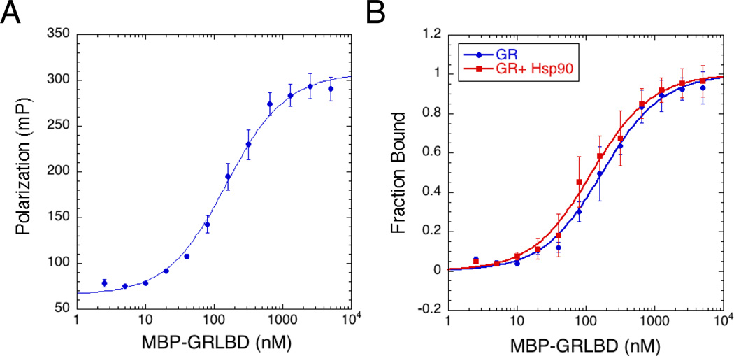
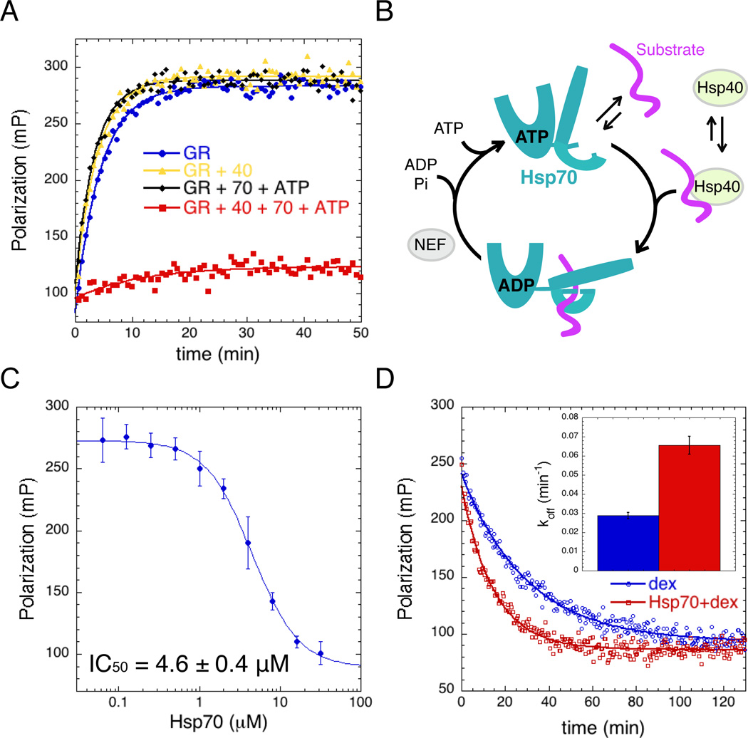
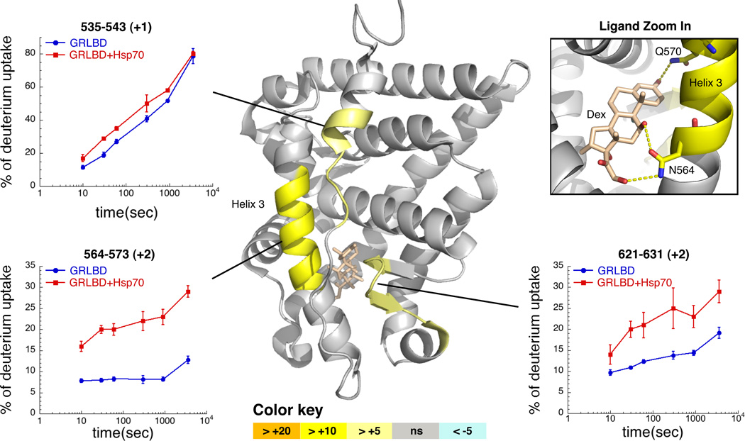
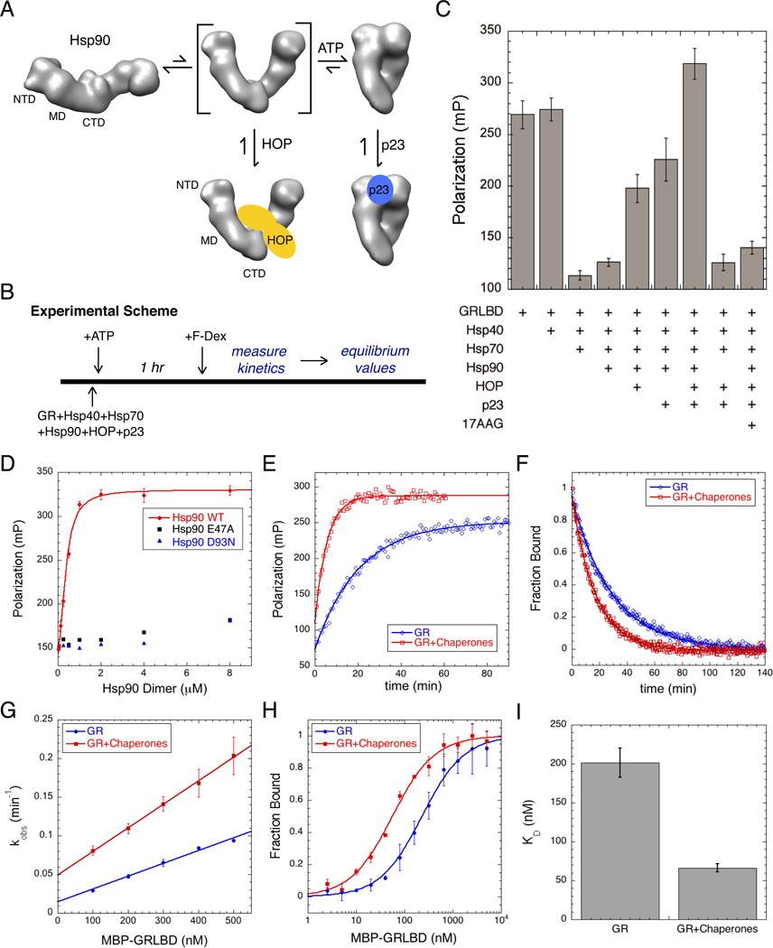
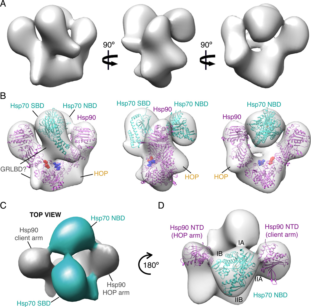
The glucocorticoid receptor (GR), like many signaling proteins, depends on the Hsp90 molecular chaperone for in vivo function. Although Hsp90 is required for ligand binding in vivo, purified apo GR is capable of binding ligand with no enhancement from Hsp90. We reveal that Hsp70, known to facilitate client delivery to Hsp90, inactivates GR through partial unfolding, whereas Hsp90 reverses this inactivation. Full recovery of ligand binding requires ATP hydrolysis on Hsp90 and the Hop and p23 cochaperones. Surprisingly, Hsp90 ATP hydrolysis appears to regulate client transfer from Hsp70, likely through a coupling of the two chaperone's ATP cycles. Such coupling is embodied in contacts between Hsp90 and Hsp70 in the GR:Hsp70:Hsp90:Hop complex imaged by cryoelectron microscopy. Whereas GR released from Hsp70 is aggregation prone, release from Hsp90 protects GR from aggregation and enhances its ligand affinity. Together, this illustrates how coordinated chaperone interactions can enhance stability, function, and regulation.


Figures

Figure 1
Figure 1. GR Ligand Binding Activity In Vitro Is Hsp90 Independent
A) Equilibrium binding of 20nM F-dex to GRLBD measured by fluorescence polarization (±SD). Binding curve fit a KD of 154±14nM (±SEM). B) Equilibrium binding with 5mM ATP/MgCl2. GRLBD alone (blue) and with 5µM Hsp90 (red) (±SD).
Figure 2
Figure 2. Hsp70 inhibits GR Ligand Binding
A) Association kinetics of F-dex to GRLBD (blue), with Hsp40 (yellow), with Hsp70 and ATP (black), and with Hsp40, Hsp70 and ATP (red). Assay conditions: 5mM ATP/MgCl2, 50nM F-dex, 1µM MBP-GRLBD, 2µM Hsp40 and 15µM Hsp70. B) Chaperone cycle of Hsp70. ATP bound NBD opens the SBD such that substrate binding is weak. Substrate and Hsp40 stimulate ATP hydrolysis, resulting in a high affinity substrate-bound state with the lid latched down. Substrate release occurs upon ADP:ATP exchange promoted by nucleotide exchange factors (NEF). C) Hsp70 concentration dependence of equilibrium binding of 1 µM MBP-GRLBD to 20nM F-dex (with 2µM Hsp40) (±SD). Fitting a cooperative competitive binding model yields an IC50 of 4.6±0.4 µM and a Hill coefficient of 1.6±0.4 µM (±SEM). D) Dissociation of 100nM F-dex from MBP-GRLBD (with 2µM Hsp40) initiated with excess (100µM) unlabeled dex (blue), and with 15µM Hsp70 (red), fit to a single exponential decay. Inset shows average off rate; 0.029±0.002 min−1 and 0.066±0.005 min−1 with Hsp70 (±SEM). See also Figure S1.
Figure 3
Figure 3. Hydrogen Exchange Mass Spectrometry Detects Partial Unfolding of GRLBD by Hsp70
Differential HDX between GRLBD and Hsp70 bound GRLBD mapped onto the dex and coactivator peptide (not shown) bound crystal structure of GRLBD (1M2Z). Differences in the average HDX are represented as percentage change and colored according to the key with orange being faster with Hsp70. Number within parentheses is the standard deviation between 3 replicates. Corresponding deuterium build-up curves for the regions that undergo the most significant conformational change are shown (±SD). Top right shows zoom in on dex, and the three hydrogen bonds formed with helix 3. See also Figure S2.
Figure 4
Figure 4. Hsp90 System Recovers GR Ligand Binding From Hsp70 Inhibition and Enhances Ligand Association
A) Without nucleotide, Hsp90 is in an extended open state. Rotation of the NTD about the MD interface is required for dimerization of the NTDs in the ATP stabilized closed state. HOP binds the intermediate open state with rotated NTDs, and p23 binds the closed ATP bound state. B) Experimental scheme for Figure 4. C) Equilibrium binding of 20nM F-dex to 1 µM MBP-GRLBD with different chaperone components (±SD). Assay conditions; 50µM 17AAG, 2µM Hsp40, and 15µM Hsp70, Hsp90, HOP, and p23. D) Saturation plot for binding of 20nM F-dex to 1 µM MBP-GRLBD with 2µM Hsp40, and 15µM Hsp70, HOP and p23, with increasing Hsp90 WT (red), and hydrolysis dead Hsp90; E47A (black) and D93N (blue) (±SD). WT Hsp90 binding curve fit to a half maximal effective concentration equation. E) Association kinetics of 20nM F-dex to 300nM MBP-GRLBD alone (blue) and with 2µM Hsp40, and 15µM Hsp70, Hsp90, HOP, and p23 (red), fit to a single phase association. F) Normalized dissociation kinetics of 100nM F-dex bound to 1µM MBP-GRLBD (blue) and with 15µM Hsp70, 2µM Hsp40, and 10µM Hsp90, HOP, and p23 (red). Off rates are respectively 0.041 ±0.004 min−1 and 0.059±0.002 min−1 (±SEM) (Figure S4D). G) Average kobs vs GRLBD concentration from 3–5 separate experiments for GRLBD (blue) and with 15µM Hsp70, 2µM Hsp40, and 10µM Hsp90, HOP, and p23 (red) (±SEM). On rates determined from the slope of the linear fit to be 0.165±0.008 and 0.304±0.072 µM−1min−1 with chaperones (± weighted error of slope) (Figure S4C). H) Normalized equilibrium binding of 20nM F-dex to GRLBD alone (blue) and with 15µM Hsp70, 2µM Hsp40, and 10µM Hsp90, HOP, and p23 (red) averaged from 2 separate experiments (±SD). I) Average ligand KD for GRLBD with and without chaperones determined from 5 separate experiments (as in Figure 4H). KD decreases from 201±42nM to 66±12nM with chaperones (±SEM). See also Figure S3 and S4.
Figure 5
Figure 5. Cryo-EM Reconstruction of Hsp90, Hsp70, HOP, GRLBD Complex
A) Cryo-EM reconstruction of Hsp90, Hsp70, HOP, GRLBD complex filtered to 38Å. B) Cryo-EM reconstruction as in A with placement of Hsp70 NTD (3ATU) and SBD (1DKX) in cyan, and Hsp90 NTD rotated HOP model (Southworth and Agard, 2011) in magenta. Client binding site residues for E. coli Hsp90; E466, W467, N470 shown in red and M546, M550, L553, F554 in blue (Genest et al., 2013). C) Top view shows density for the linker between Hsp70 domains (cyan). D) Top view showing positioning of Hsp70 NTD between Hsp90 NTDs. See also Figure S5 and S6.
Figure 6
Figure 6. Hydrolysis Independent Chaperone Function of Hsp90 is Important for Maintaining an Active GR Population
A) Experimental scheme for B. GRLBD was pre-inhibited by Hsp70 and Hsp40, and equilibrated with F-dex. Ligand binding is initiated with either Bag-1 or the Hsp90 system. B) Ligand binding initiated with,Bag-1 (black), Hsp90 with HOP and p23 (red), Hsp90(E47A) with HOP and p23 (green), Bag-1 plus Hsp90 with HOP and p23 (purple), and Bag-1 with Hsp90(E47A) with HOP and p23 (yellow). Also shown, GRLBD alone preincubated with F-dex (blue). Assay conditions: 50nM F-dex, 1µM MBP-GRLBD, 2µM Hsp40, 15µM Hsp70, Hsp90, HOP, p23 and Bag-1. C) Time course of light scattering for GRLBD (blue), and for GRLBD preincubated with Hsp40 and Hsp70 as for (A), with time course initiated with Bag-1 (black). Same conditions as B but without F-dex. See also Figure S7.
Figure 7
Figure 7. Model for GR Ligand Binding
Apo GRLBD on its own is mostly folded and transiently samples a more unstructured state where the binding pocket at the core of the protein is accessible, allowing ligand association and dissociation. In a process that requires ATP hydrolysis and Hsp40, Hsp70 binds to GRLBD and promotes ligand release by stabilizing a partially unfolded inactive state. When GRLBD release from Hsp70 is stimulated by the NEF, Bag-1, GRLBD is rapidly released in a partially unfolded state that while able to bind ligand, is prone to aggregation. In the presence of Hsp90, GRLBD bound Hsp70 is brought together with Hsp90 by HOP, to form an inactive complex. Subsequent ATP hydrolysis on Hsp90 is required to release Hsp70 (and HOP), allowing progression to the closed state, and p23 binding. Together, Hsp90 and Bag-1 cooperate to promote Hsp70 release such that Hsp90 ATP hydrolysis is not required. Progression to the matured complex with Hsp90 and p23 results in high affinity ligand binding.

References

    1. Ali MMU, Roe SM, Vaughan CK, Meyer P, Panaretou B, Piper PW, Prodromou C, Pearl LH. Crystal structure of an Hsp90–nucleotide–p23/Sba1 closed chaperone complex. Nature. 2006;440:1013–1017. - PMC - PubMed
    1. Bain DL, Heneghan AF, Connaghan-Jones KD, Miura MT. Nuclear Receptor Structure: Implications for Function. Annu. Rev. Physiol. 2007;69:201–220. - PubMed
    1. Bledsoe RK, Montana VG, Stanley TB, Delves CJ, Apolito CJ, McKee DD, Consler TG, Parks DJ, Stewart EL, Willson TM. Crystal Structure of the Glucocorticoid Receptor Ligand Binding Domain Reveals a Novel Mode of Receptor Dimerization and Coactivator Recognition. Cell. 2002;110:93–105. - PubMed
    1. Chen S, Smith DF. Hop as an adaptor in the heat shock protein 70 (Hsp70) and hsp90 chaperone machinery. J. Biol. Chem. 1998;273:35194–35200. - PubMed
    1. Chrousos GP, Kino T. Glucocorticoid Signaling in the Cell. Annals of the New York Academy of Sciences. 2009;1179:153–166. - PMC - PubMed

Publication types

MeSH terms

Substances