Review
. 2014 Aug;37(8):443-54.
doi: 10.1016/j.tins.2014.05.005. Epub 2014 Jun 17.

From molecule to mind: an integrative perspective on odor intensity

Affiliations
Review

From molecule to mind: an integrative perspective on odor intensity

Joel D Mainland et al. Trends Neurosci. 2014 Aug.

Abstract

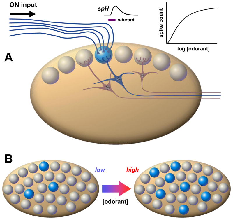
A fundamental problem in systems neuroscience is mapping the physical properties of a stimulus to perceptual characteristics. In vision, wavelength translates into color; in audition, frequency translates into pitch. Although odorant concentration is a key feature of olfactory stimuli, we do not know how concentration is translated into perceived intensity by the olfactory system. A variety of neural responses at several levels of processing have been reported to vary with odorant concentration, suggesting specific coding models. However, it remains unclear which, if any, of these phenomena underlie the perception of odor intensity. Here, we provide an overview of current models at different stages of olfactory processing, and identify promising avenues for future research.


Figures

Figure I
Figure I
Figure 1
Figure 1. Odorant concentration coding in olfactory sensory neurons (OSNs)
During sensory transduction (A), odorant molecules bind and stabilize the active states of olfactory receptors (R) in ciliary membranes of OSNs. The activated receptors (R*) couple to G-proteins (Golf) and increase synthesis of cyclic AMP (cAMP) by type III adenylyl cyclase (AC3). The cAMP opens cyclic nucleotide-gated channels that conduct calcium ions into the cilia and in turn open a channel (ANO2) mediating a depolarizing efflux of chloride ions. The resulting transduction current is passed to the OSN cell body where it drives a train of action potentials (spikes). The concentration of detected odorant is encoded non-linearly at each step of transduction: by a hyperbolic dependence on the number of activated receptors (R*) in the cilia (B), a strongly cooperative variation in amplitude of the transduction current (C), and similar sigmoidal variation of spike firing rate relayed by OSN axons (D). Data source: C, D: [115], normalized currents and firing rates of frog OSN response to cineole; mammalian OSNs exhibit similar dose-response profiles.
Figure 2
Figure 2. Spatial and temporal coding of odorant concentration in the olfactory bulb
A. Schematic of four parallel glomerular odor-encoding channels. During responses to odorant stimuli sampled by a single sniff, olfactory nerve (ON) spike inputs to different glomeruli exhibit different latencies and time courses (monitored by calcium indicator fluorescence signals). This reflects the diversity of olfactory receptor molecular tunings. These spatiotemporal patterns of input are processed by glomerular neural circuits, including excitatory external tufted (ET), as well as inhibitory periglomerular (PG) and short axon (SA) cells, before being encoded into spiking patterns of mitral cells (MC) for relay to olfactory cortex. The SA cells have widespread interglomerular projections that could act to normalize spatial patterns of glomerular output as input patterns vary with odorant concentration (c.f. Fig. 5B). A second layer of signal filtering and pattern transformation occurs when granule cells (GC) or other local interneurons inhibit mitral cells. B. As odorant concentration is increased from low (blue) to high (red), OSNs are activated more rapidly and presynaptic glomerular responses have shorter latencies (c.f. red ON traces). Mitral cell spike output may track these latency shifts over the sniff cycle (c.f. left-shifted red spike responses in glomerular channels 1, 3 & 4). Such phase shifts of spike latency over sniff cycles may encode odor concentration, while leaving invariant overall patterns of relative latencies between glomeruli (which can encode odor quality, as latencies will vary with different sensitivities of receptors to different odorants). This simple scheme allows early separation of the coding of odor concentration vs. odor quality, but may be complicated by lateral inhibition between glomeruli. Some mitral cells exhibit suppressed basal spiking in response to an odor, which may be followed by rebound spiking whose latency becomes longer as odorant concentration is increased (due to stronger inhibition) (as pictured in glomerular channel 2). C. Example of excitatory mitral cell response in a glomerular coding channel. Data show a logarithmic decrement of mitral cell spike latency with increasing concentration, which provides an immediate readout of intensity (upper panel). Mitral cell spike count and mean spike rate over a sniff (middle and lower panels) increase, so spike rate coding of concentration is possible if readouts have longer integration times. Data sources: C: upper panel: [116], salamander mitral/ tufted cell excitatory response to isoamyl acetate; middle panel: [41], mean spikes per sniff cycle for rat mitral/ tufted cell response to cineole; lower panel: [117], mouse mitral/ tufted cell spike response evoked by heptanal activation of olfactory receptor I7 (mean of 8 cells, spike rate increment over baseline rate, normalized to maximum rate).
Figure 3
Figure 3. Odorant concentration coding in glomerular circuits
A. Each glomerulus transforms the afferent ON signals transduced by one type of olfactory receptor into the spike activities of several dozen or more mitral cells (MC) and tufted cells (TC). The two classes (MC, TC) of olfactory bulb output neurons differ in connectivity and regulation by local circuits. Various subtypes of tufted cells are located more superficially, receive more direct ON input and less granule cell inhibition, and are more easily excited. The mitral cells are located deeper, receive mostly indirect ON input (via ET cells) and more granule cell inhibition, and have higher spike thresholds. B. Coding of odorant concentration by the number or fraction of responding ‘sister’ mitral/ tufted (i.e. connected to the same glomerulus). When odorant concentration is increased, stronger afferent ON input to the glomerulus can recruit a greater proportion of sister mitral/ tufted cells, including those with higher spike thresholds. C. Coding of odorant concentration by shift in spike latency between mitral and tufted cells. When odorant concentration is increased (blue to red traces), latency and sniff cycle phase of mitral cell spike responses decreases while that of tufted cells remains approximately invariant. D. Coding of odorant concentration by medio-lateral timing difference in activation of pairs of glomeruli corresponding to the same olfactory receptor. Inhaled odorant gains access to OSNs in medial olfactory epithelium (on septum) sooner than OSNs in lateral olfactory epithelium (in recesses of nasal turbinates). This results in a latency difference between inputs to glomeruli of mirror image receptor maps in medial and lateral halves of the olfactory bulb. E. Medio-lateral gradient in concentration at fixed time due to relative lag in odorant access. This gradient may depend on sorption properties of the odorant. F. As odorant concentration is increased, the latency of spike responses drops at different rates for mitral cells connected to medial vs. lateral glomeruli. Data sources: C: [46], average spike phase of mouse mitral cell vs. tufted cell response to various odorants; F: [49], onset latencies of mitral cell spike responses in medial vs. lateral olfactory bulb of transgenic mice with expression of olfactory receptor I7 in all OSNs activated by octanal.
Figure 4
Figure 4. Odorant concentration coding in olfactory bulb glomeruli
A. Thousands of OSNs expressing the same olfactory receptor relay convergent synaptic input via the olfactory nerve (ON) to one or a few glomeruli at stereotypic locations in the olfactory bulb. The collective inputs to individual glomeruli can be quantified by optical measurements of odorant responses using presynaptic indicators of calcium signaling, or transmitter release (e.g. the exocytosis reporter synaptopHluorin, spH). This reveals coding of odorant concentration by a weakly cooperative dose- response curve. B. Each glomerulus receives input from one type of receptor out of a diverse population of ~ 102 – 103 different receptors, with different odorant tunings. This means that as concentration of a particular odorant increases, a large set of receptors (and hence glomeruli) is recruited. This spatial expansion of the number of activated glomeruli (shown here in blue) has been suggested to serve as a concentration code at the system level.

References

    1. Chastrette M, et al. Modelling the human olfactory stimulus-response function. Chem Senses. 1998;23:181–196. - PubMed
    1. Henion KE. Odor pleasantness and intensity: a single dimension? J Exp Psychol. 1971;90:275–279. - PubMed
    1. Gross-Isseroff R, Lancet D. Concentration-dependent changes of perceived odor quality. Chem Senses. 1988;13:191.
    1. Wojcik PT, Sirotin YB. Single scale for odor intensity in rat olfaction. Current biology : CB. 2014;24:568–573. - PubMed
    1. Eichenbaum H, et al. Selective olfactory deficits in case H.M. Brain. 1983;106 (Pt 2):459–472. - PubMed

Publication types

LinkOut - more resources