

Atorvastatin helps preserve pancreatic β cell function in obese C57BL/6 J mice and the effect is related to increased pancreas proliferation and amelioration of endoplasmic-reticulum stress
- PMID: 24950764
- PMCID: PMC4078942
- DOI: 10.1186/1476-511X-13-98
Atorvastatin helps preserve pancreatic β cell function in obese C57BL/6 J mice and the effect is related to increased pancreas proliferation and amelioration of endoplasmic-reticulum stress
Abstract
Background: 3-Hydroxy-3-methyl-glutaryl CoA (HMG-CoA) reductase inhibitors or statins are competitive inhibitors of the rate-limiting enzyme in cholesterol biosynthesis. Currently, statins are used as first-line therapy in the treatment of diabetic dyslipidemia. However, effects of statins on β cell function remains unclear. This study aims to examine effects of atorvastatin treatment on pancreatic β cell function in obese C57BL/6 J mice and the possible mechanisms.
Methods: Diet-induced obesity (DIO) C57BL/6 J mice were treated with atorvastatin (30 mg/kg/day) for 58 days. β cell function was assessed by hyperglycemic clamp and the area of insulin-positive β cells was examined by immunofluorescence. Gene expression was assessed by RT-PCR, and endoplasmic reticulum (ER) stress related proteins were examined by Western blot. Additionally, cell viability and apoptosis of the cholesterol-loaded NIT-1 cells were investigated after atorvastatin treatment.
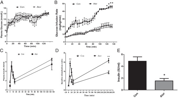
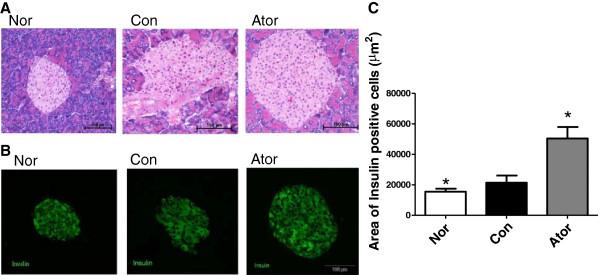
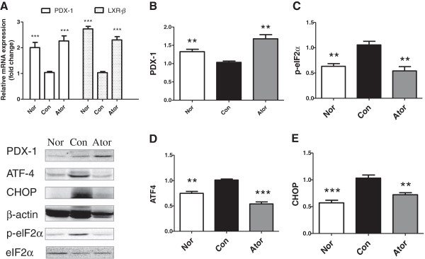
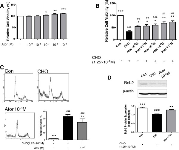
Results: Hyperglycemic clamp study revealed that glucose infusion rate (GIR) and insulin stimulation ratio in atorvastatin-treated DIO mice were markedly higher than control mice (P < 0.05, P < 0.01 vs. con), indicating preserved β-cell sensitivity to glucose. Lipid profiles of plasma triglyceride (TG), pancreas TG and plasma cholesterol (CHO) were improved. Pancreas weight and weight index were improved significantly after atorvastatin treatment (P < 0.05 vs. con). Immunofluorescence results showed that atorvastatin-treated mice had significantly larger insulin-positive β cell area (P < 0.05 vs. con). Furthermore, RT-PCR and western blot showed that the mRNA and protein expression of pancreatic and duodenal homeobox 1 (Pdx1) in the pancreas were upregulated (P < 0.001, P < 0.01 vs. con). Moreover, the expression level of ER stress markers of activating transcription factor 4 (ATF4), CCAAT-enhancer-binding protein homologous protein (CHOP) and phosphorylated eukaryotic initiation factor 2α (eIF2α) were downregulated in the pancreas of atorvastatin-treated mice (P < 0.001, P < 0.01, P < 0.01 vs. con). Besides, atorvastatin protected the pancreatic β cell line of NIT-1 from cholesterol-induced apoptosis. Western blot showed increased expression of anti-apoptotic protein of B-cell lymphoma 2 (Bcl-2).
Conclusion: Pancreatic β cell function of obese C57BL/6 J mice was preserved after atorvastatin treatment, and this improvement may be attributed to enhanced pancreas proliferation and amelioration of pancreatic ER stress.
Figures
Similar articles
-
A2a adenosine receptor agonist improves endoplasmic reticulum stress in MIN6 cell line through protein kinase A/ protein kinase B/ Cyclic adenosine monophosphate response element-binding protein/ and Growth Arrest And DNA-Damage-Inducible 34/ eukaryotic Initiation Factor 2α pathways.J Cell Physiol. 2019 Jul;234(7):10500-10511. doi: 10.1002/jcp.27719. Epub 2018 Nov 11. J Cell Physiol. 2019. PMID: 30417358
-
Endoplasmic reticulum stress contributes to NMDA-induced pancreatic β-cell dysfunction in a CHOP-dependent manner.Life Sci. 2019 Sep 1;232:116612. doi: 10.1016/j.lfs.2019.116612. Epub 2019 Jun 28. Life Sci. 2019. PMID: 31260687
-
Emulsified isoflurane protects beta cells against high glucose-induced apoptosis via inhibiting endoplasmic reticulum stress.Ann Palliat Med. 2020 Jan;9(1):90-97. doi: 10.21037/apm.2019.11.31. Ann Palliat Med. 2020. PMID: 32005067
-
Atorvastatin delays progression of pancreatic lesions to carcinoma by regulating PI3/AKT signaling in p48Cre/+ LSL-KrasG12D/+ mice.Int J Cancer. 2012 Oct 15;131(8):1951-62. doi: 10.1002/ijc.27456. Epub 2012 Mar 14. Int J Cancer. 2012. PMID: 22287227 Free PMC article.
-
Endoplasmic reticulum stress and eIF2α phosphorylation: The Achilles heel of pancreatic β cells.Mol Metab. 2017 Jul 12;6(9):1024-1039. doi: 10.1016/j.molmet.2017.06.001. eCollection 2017 Sep. Mol Metab. 2017. PMID: 28951826 Free PMC article. Review.
Cited by
-
Dual Effect of Rosuvastatin on Glucose Homeostasis Through Improved Insulin Sensitivity and Reduced Insulin Secretion.EBioMedicine. 2016 Aug;10:185-94. doi: 10.1016/j.ebiom.2016.07.007. Epub 2016 Jul 9. EBioMedicine. 2016. PMID: 27453321 Free PMC article.
-
The additive effects of atorvastatin and insulin on renal function and renal organic anion transporter 3 function in diabetic rats.Sci Rep. 2017 Oct 19;7(1):13532. doi: 10.1038/s41598-017-13206-5. Sci Rep. 2017. PMID: 29051569 Free PMC article.
-
Therapeutic Effect of Adipose Derived Stem Cells versus Atorvastatin on Amiodarone Induced Lung Injury in Male Rat.Int J Stem Cells. 2015 Nov;8(2):170-80. doi: 10.15283/ijsc.2015.8.2.170. Int J Stem Cells. 2015. PMID: 26634065 Free PMC article.
-
Pancreatic Steatosis as a Risk Factor for Pancreatic Ductal Adenocarcinoma: Pathogenesis and Clinical Implications.Clin Transl Gastroenterol. 2025 Feb 24;16(6):e00832. doi: 10.14309/ctg.0000000000000832. eCollection 2025 Jun 1. Clin Transl Gastroenterol. 2025. PMID: 39991930 Free PMC article. Review.
-
Increases in bioactive lipids accompany early metabolic changes associated with β-cell expansion in response to short-term high-fat diet.Am J Physiol Endocrinol Metab. 2018 Dec 1;315(6):E1251-E1263. doi: 10.1152/ajpendo.00001.2018. Epub 2018 Aug 14. Am J Physiol Endocrinol Metab. 2018. PMID: 30106624 Free PMC article.
References
-
- Mason RP, Walter MF, Day CA, Jacob RF. Intermolecular differences of 3-hydroxy-3-methylglutaryl coenzyme a reductase inhibitors contribute to distinctpharmacologic and pleiotropic actions. Am J Cardiol. 2005;96:11–23. - PubMed
-
- Heart Protection Study Collaborative Group. MRC/BHF Heart Protection Study of cholesterol lowering with simvastatin in 20,536 high-risk individuals: a randomised placebo-controlled trial. Lancet. 2002;360:7–22. - PubMed
-
- Lehto S, Rönnemaa T, Haffner SM, Pyörälä K, Kallio V, Laakso M. Dyslipidemia and hyperglycemia predict coronary heart disease events in middle-aged patients with NIDDM. Diabetes. 1997;46:1354–1359. - PubMed
-
- Grundy SM, Cleeman JI, Merz CN, Brewer HB Jr, Clark LT, Hunninghake DB, Pasternak RC, Smith SC Jr, Stone NJ. National Heart, Lung, and Blood Institute; American College of Cardiology Foundation; American Heart Association. Implications of recent clinical trials for the National Cholesterol Education Program Adult Treatment Panel III guidelines. Circulation. 2004;110:227–239. - PubMed
Publication types
MeSH terms
Substances
LinkOut - more resources
Full Text Sources
Other Literature Sources
Research Materials
Miscellaneous
