

ERM proteins at a glance
- PMID: 24951115
- PMCID: PMC4117225
- DOI: 10.1242/jcs.098343
ERM proteins at a glance
Abstract
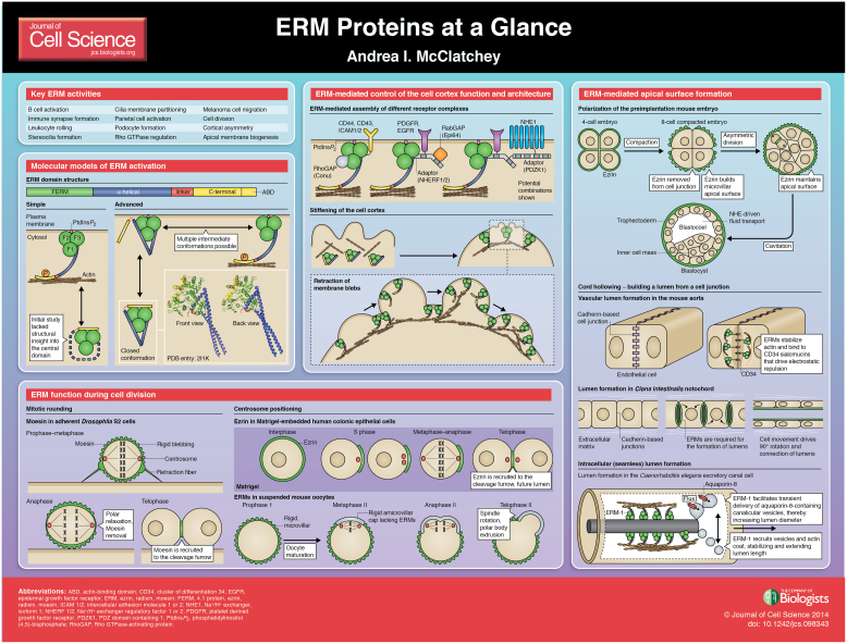
The cell cortex is a dynamic and heterogeneous structure that governs cell identity and behavior. The ERM proteins (ezrin, radixin and moesin) are major architects of the cell cortex, and they link plasma membrane phospholipids and proteins to the underlying cortical actin cytoskeleton. Recent studies in several model systems have uncovered surprisingly dynamic and complex molecular activities of the ERM proteins and have provided new mechanistic insight into how they build and maintain cortical domains. Among many well-established and essential functions of ERM proteins, this Cell Science at a Glance article and accompanying poster will focus on the role of ERMs in organizing the cell cortex during cell division and apical morphogenesis. These examples highlight an emerging appreciation that the ERM proteins both locally alter the mechanical properties of the cell cortex, and control the spatial distribution and activity of key membrane complexes, establishing the ERM proteins as a nexus for the physical and functional organization of the cell cortex and making it clear that they are much more than scaffolds. This article is part of a Minifocus on Establishing polarity.
Keywords: Actin cytoskeleton; Apical morphogenesis; Cell cortex; Ezrin; Moesin; Radixin.
© 2014. Published by The Company of Biologists Ltd.
Comment on
-
Establishment of epithelial polarity--GEF who's minding the GAP?J Cell Sci. 2014 Aug 1;127(Pt 15):3205-15. doi: 10.1242/jcs.153197. Epub 2014 Jul 2. J Cell Sci. 2014. PMID: 24994932 Free PMC article. Review.
-
Integrins and epithelial cell polarity.J Cell Sci. 2014 Aug 1;127(Pt 15):3217-25. doi: 10.1242/jcs.146142. Epub 2014 Jul 2. J Cell Sci. 2014. PMID: 24994933 Free PMC article. Review.
References
-
- Brown M. J., Nijhara R., Hallam J. A., Gignac M., Yamada K. M., Erlandsen S. L., Delon J., Kruhlak M., Shaw S. (2003). Chemokine stimulation of human peripheral blood T lymphocytes induces rapid dephosphorylation of ERM proteins, which facilitates loss of microvilli and polarization. Blood 102, 3890–3899 10.1182/blood-2002-12-3807 - DOI - PubMed
Publication types
MeSH terms
Substances
LinkOut - more resources
Full Text Sources
Other Literature Sources
Research Materials
