

Adhesion of Staphylococcus aureus to the vessel wall under flow is mediated by von Willebrand factor-binding protein
- PMID: 24951431
- PMCID: PMC7025350
- DOI: 10.1182/blood-2014-02-558890
Adhesion of Staphylococcus aureus to the vessel wall under flow is mediated by von Willebrand factor-binding protein
Abstract
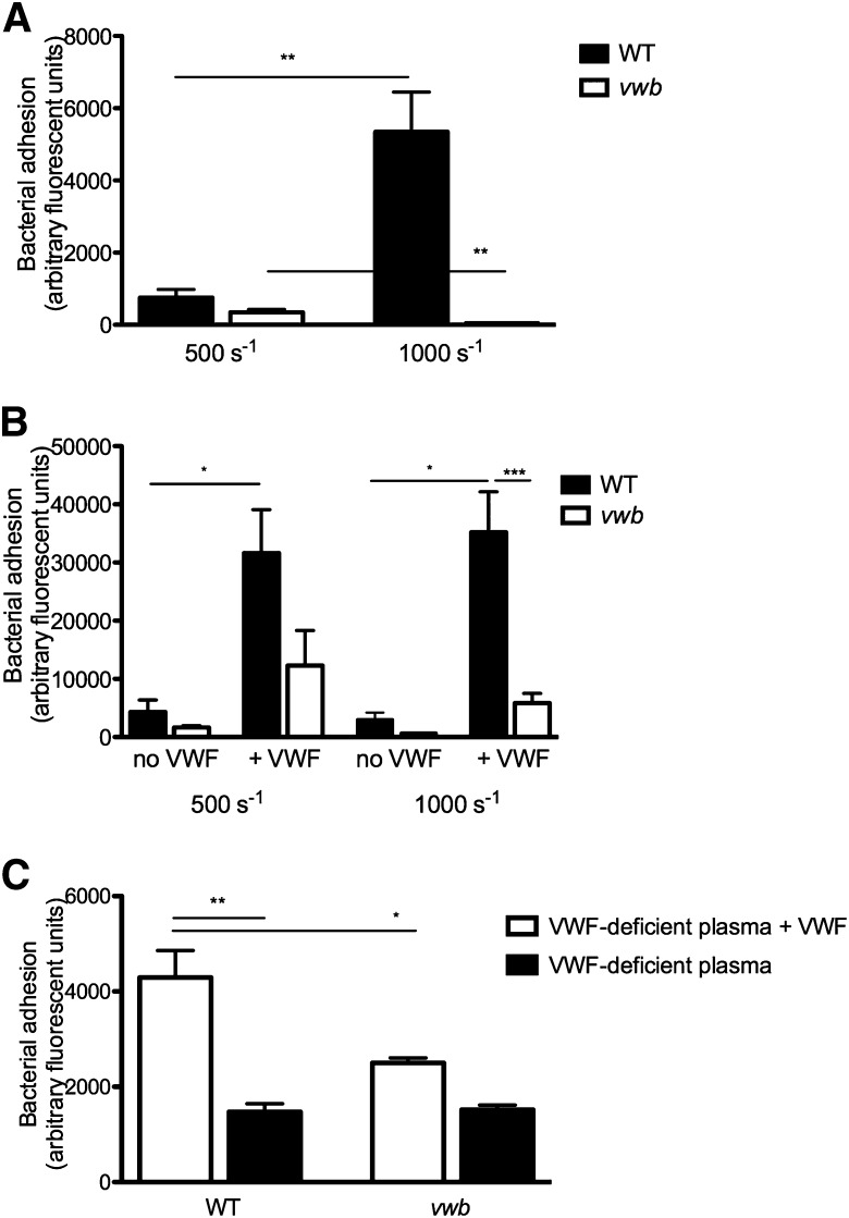
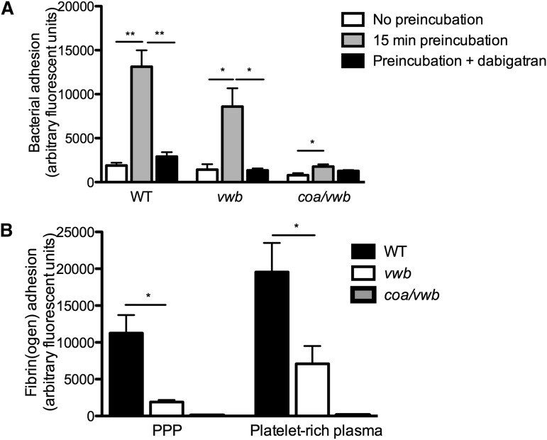
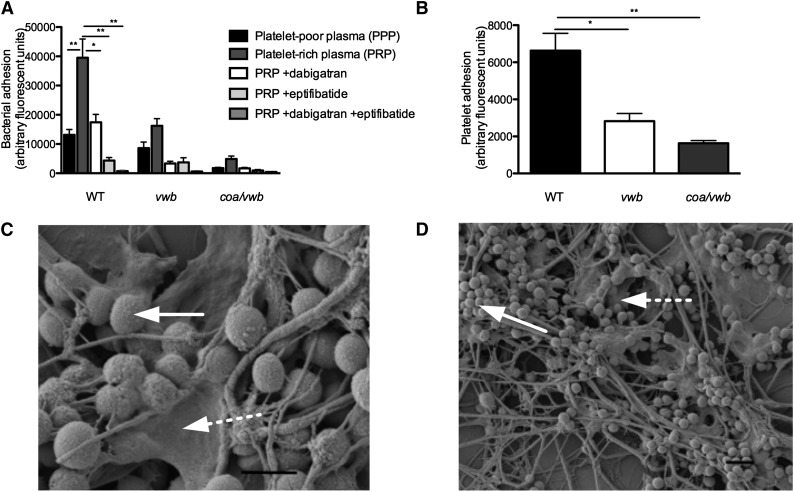
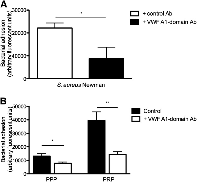
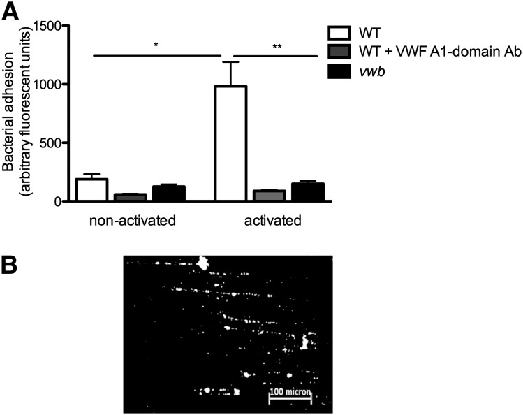
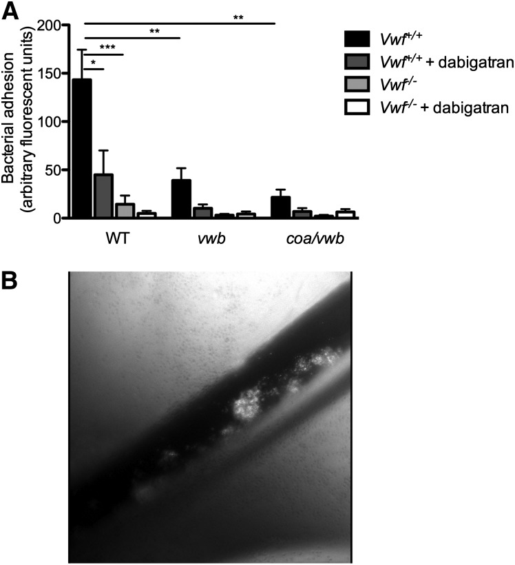
Adhesion of Staphylococcus aureus to blood vessels under shear stress requires von Willebrand factor (VWF). Several bacterial factors have been proposed to interact with VWF, including VWF-binding protein (vWbp), a secreted coagulase that activates the host's prothrombin to generate fibrin. We measured the adhesion of S aureus Newman and a vWbp-deficient mutant (vwb) to VWF, collagen, and activated endothelial cells in a microparallel flow chamber. In vivo adhesion of S aureus was evaluated in the mesenteric circulation of wild-type (WT) and VWF-deficient mice. We found a shear-dependent increase in adhesion of S aureus to the (sub)endothelium that was dependent on interactions between vWbp and the A1-domain of VWF. Adhesion was further enhanced by coagulase-mediated fibrin formation that clustered bacteria and recruited platelets into bacterial microthrombi. In vivo, deficiency of vWbp or VWF as well as inhibition of coagulase activity reduced S aureus adhesion. We conclude that vWbp contributes to vascular adhesion of S aureus through 2 independent mechanisms: shear-mediated binding to VWF and activation of prothrombin to form S aureus-fibrin-platelet aggregates.
© 2014 by The American Society of Hematology.
Conflict of interest statement
Conflict-of-interest disclosure: P.V. has received honoraria from Boehringer-Ingelheim for lectures and advisory committees. The remaining authors declare no competing financial interests.
Figures
Comment in
-
Go with the flow: S aureus and vascular infection.Blood. 2014 Sep 4;124(10):1549-51. doi: 10.1182/blood-2014-07-583864. Blood. 2014. PMID: 25190752 Free PMC article.
Similar articles
-
Clumping factor A, von Willebrand factor-binding protein and von Willebrand factor anchor Staphylococcus aureus to the vessel wall.J Thromb Haemost. 2017 May;15(5):1009-1019. doi: 10.1111/jth.13653. Epub 2017 Mar 23. J Thromb Haemost. 2017. PMID: 28182324 Free PMC article.
-
Ultralarge von Willebrand factor fibers mediate luminal Staphylococcus aureus adhesion to an intact endothelial cell layer under shear stress.Circulation. 2013 Jul 2;128(1):50-9. doi: 10.1161/CIRCULATIONAHA.113.002008. Epub 2013 May 29. Circulation. 2013. PMID: 23720451
-
The Expression of von Willebrand Factor-Binding Protein Determines Joint-Invading Capacity of Staphylococcus aureus, a Core Mechanism of Septic Arthritis.mBio. 2020 Nov 17;11(6):e02472-20. doi: 10.1128/mBio.02472-20. mBio. 2020. PMID: 33203754 Free PMC article.
-
Platelet-vessel wall interactions in thrombosis and restenosis role of von Willebrand factor.Verh K Acad Geneeskd Belg. 1997;59(3):161-83. Verh K Acad Geneeskd Belg. 1997. PMID: 9490916 Review.
-
[Von Willebrand factor and platelet adhesion to the subendothelium of the vascular wall].Nouv Rev Fr Hematol (1978). 1982;24(3):145-51. Nouv Rev Fr Hematol (1978). 1982. PMID: 6752879 Review. French.
Cited by
-
Native valve, prosthetic valve, and cardiac device-related infective endocarditis: A review and update on current innovative diagnostic and therapeutic strategies.Front Cell Dev Biol. 2022 Oct 3;10:995508. doi: 10.3389/fcell.2022.995508. eCollection 2022. Front Cell Dev Biol. 2022. PMID: 36263017 Free PMC article. Review.
-
Novel dalbavancin-PLLA implant coating prevents hematogenous Staphylococcus aureus infection in a minimally invasive mouse tail vein model.Front Bioeng Biotechnol. 2022 Nov 17;10:1021827. doi: 10.3389/fbioe.2022.1021827. eCollection 2022. Front Bioeng Biotechnol. 2022. PMID: 36466340 Free PMC article.
-
Exploring the role of bacterial virulence factors and host elements in septic arthritis: insights from animal models for innovative therapies.Front Microbiol. 2024 Feb 12;15:1356982. doi: 10.3389/fmicb.2024.1356982. eCollection 2024. Front Microbiol. 2024. PMID: 38410388 Free PMC article. Review.
-
Effect of Gravity on Bacterial Adhesion to Heterogeneous Surfaces.Pathogens. 2023 Jul 15;12(7):941. doi: 10.3390/pathogens12070941. Pathogens. 2023. PMID: 37513788 Free PMC article.
-
Binding of Staphylococcus aureus Protein A to von Willebrand Factor Is Regulated by Mechanical Force.mBio. 2019 Apr 30;10(2):e00555-19. doi: 10.1128/mBio.00555-19. mBio. 2019. PMID: 31040240 Free PMC article.
References
-
- Vanassche T, Peetermans WE, Herregods MC, Herijgers P, Verhamme P. Anti-thrombotic therapy in infective endocarditis. Expert Rev Cardiovasc Ther. 2011;9(9):1203–1219. - PubMed
-
- Kern WV. Management of Staphylococcus aureus bacteremia and endocarditis: progresses and challenges. Curr Opin Infect Dis. 2010;23(4):346–358. - PubMed
-
- Corey GR. Staphylococcus aureus bloodstream infections: definitions and treatment. Clinical Infect Dis. 2009;48(Suppl 4):S254–259. - PubMed
-
- Bjerketorp J, Jacobsson K, Frykberg L. The von Willebrand factor-binding protein (vWbp) of Staphylococcus aureus is a coagulase. FEMS Microbiol Lett. 2004;234(2):309–314. - PubMed
-
- Sadler JE. Biochemistry and genetics of von Willebrand factor. Annu Rev Biochem. 1998;67:395–424. - PubMed
Publication types
MeSH terms
Substances
LinkOut - more resources
Full Text Sources
Other Literature Sources
Molecular Biology Databases
Miscellaneous
