. 2014 Aug;20(8):1257-71.
doi: 10.1261/rna.044503.114. Epub 2014 Jun 20.

Characterization of two homologous 2'-O-methyltransferases showing different specificities for their tRNA substrates

Affiliations

Characterization of two homologous 2'-O-methyltransferases showing different specificities for their tRNA substrates

Jonathan Somme et al. RNA. 2014 Aug.

Abstract

The 2'-O-methylation of the nucleoside at position 32 of tRNA is found in organisms belonging to the three domains of life. Unrelated enzymes catalyzing this modification in Bacteria (TrmJ) and Eukarya (Trm7) have already been identified, but until now, no information is available for the archaeal enzyme. In this work we have identified the methyltransferase of the archaeon Sulfolobus acidocaldarius responsible for the 2'-O-methylation at position 32. This enzyme is a homolog of the bacterial TrmJ. Remarkably, both enzymes have different specificities for the nature of the nucleoside at position 32. While the four canonical nucleosides are substrates of the Escherichia coli enzyme, the archaeal TrmJ can only methylate the ribose of a cytidine. Moreover, the two enzymes recognize their tRNA substrates in a different way. We have solved the crystal structure of the catalytic domain of both enzymes to gain better understanding of these differences at a molecular level.

Keywords: SPOUT; methyltransferase; modified nucleosides; tRNA.


Figures

FIGURE 1.
FIGURE 1.
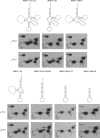
Purified Saci_0621 catalyzes the formation of Cm at position 32 of tRNA in vitro. Autoradiograms of two-dimensional chromatograms of P1 hydrolysates of [α32P]CTP-labeled transcripts of wild-type tRNAiMet of S. acidocaldarius (A) and the C32U mutant (B) incubated 30 min at 60°C in presence or in absence of purified Saci_0621. Circles in dotted lines show the migration of the pA, pG, and pU nucleotides used as UV markers. Solvent B was used for the second dimension of the chromatography. The arrows indicate the direction of migration, while the numbers indicate the order of migrations. (C) Secondary structure of tRNAiMet of S. acidocaldarius. The C32U mutation is indicated.
FIGURE 2.
FIGURE 2.
SaTrmJ shows a narrower specificity for the type of nucleoside at position 32 of tRNA than EcTrmJ. (A) Autoradiograms of two-dimensional chromatograms of T2 hydrolysates of [α32P]UTP-labeled transcripts of wild-type tRNASer of E. coli and of the C32U, C32A, and C32G mutants incubated with purified EcTrmJ or SaTrmJ at 37°C or 60°C, respectively, during 30 min. As the UmUp spot and GmUp spot comigrate with the Up spot and the Gp spot, respectively, the autoradiograms of two-dimensional chromatograms of P1 hydrolysates of [α32P]UTP-labeled transcripts of the tRNASer C32U mutant and of [α32P]GTP-labeled transcripts of the tRNASer C32G mutant incubated with purified EcTrmJ or SaTrmJ at 37°C or 60°C are shown in B. Circles in dotted lines show the migration of the pA, pG, and pC nucleotides used as UV markers. (C) Autoradiograms of two-dimensional chromatograms of T2 hydrolysates of [α32P]ATP-labeled transcripts of wild-type tRNAAla and tRNAPro of E. coli incubated with purified EcTrmJ. Solvent C was used for the second dimension of the chromatography in A, whereas solvent B was used in B and C (see Materials and Methods). The arrows indicate the direction of migration, while the numbers indicate the order of migrations.
FIGURE 3.
FIGURE 3.
Contrary to EcTrmJ, SaTrmJ is able to modify unfractionated tRNAs of E. coli. Autoradiograms of two-dimensional chromatograms of P1 hydrolysates of unfractionated tRNAs from E. coli incubated in presence of [methyl-14C]SAM and purified EcTrmJ or SaTrmJ. Circles in dotted lines show the migration of the pA, pC, pG, and pU nucleotides used as UV markers. Solvent B was used for the second dimension of the chromatography. The arrows indicate the direction of migration, while the numbers indicate the order of migrations.
FIGURE 4.
FIGURE 4.
SaTrmJ can modify truncated tRNAs, whereas EcTrmJ modifies only full-length tRNA. Secondary structures of wild-type and truncated tRNASer of E. coli are shown above the autoradiograms of two-dimensional chromatograms of T2 hydrolysates of [α32P]UTP-labeled transcripts incubated in presence of purified EcTrmJ or SaTrmJ. Solvent B was used for the second dimension of the chromatography. The arrows indicate the direction of migration, while the numbers indicate the order of migrations. For the meaning of abbreviations, see Supplemental Table S2 in the Supplemental Data.
FIGURE 5.
FIGURE 5.
Elements in the D-stem/loop are important for the tRNA recognition by EcTrmJ, whereas elements in the anticodon stem are important for the recognition by SaTrmJ. Secondary structures of the substrate E. coli tRNASer (in black), the nonsubstrate tRNAMet (in red), and hybrid tRNAs (in black and red) are shown at the left or above the autoradiograms of two-dimensional chromatograms of P1 hydrolysates of [α32P]CTP-labeled tRNA transcripts incubated in presence of purified EcTrmJ or SaTrmJ. (A) tRNASer and tRNAMet and hybrids of tRNASer with each separate stem/loop of tRNAMet. (B) tRNAMet with D- or anticodon stem/loop of tRNASer. (C) tRNASer with D-stem or -loop of tRNAMet. (D) tRNASer with anticodon stem or loop of tRNAMet. Circles in dotted lines show the migration of the pA, pG, and pU nucleotides used as UV markers. Solvent B was used for the second dimension of the chromatography. The arrows indicate the direction of migration, while the numbers indicate the order of migrations. For the meaning of abbreviations, see Supplemental Table S2 in the Supplemental Data.
FIGURE 6.
FIGURE 6.
(A) Sequence alignment of EcTrmJ and SaTrmJ. The secondary structure elements, as deduced from the crystal structures, are shown below the alignment with β-strands shown as blue arrows and α-helices as green tubes. The three predicted α-helices of the C-terminal domain are colored in orange. Identical residues in the EcTrmJ and SaTrmJ sequence are highlighted in gray. (B) Crystal structures of EcTrmJ1-164_SAH (left) and SaTrmJ1-157_SAH (right). The biological dimers are shown in cartoon representation with one subunit colored in gray. The reaction product SAH located in the active site (in purple), as well as the catalytic tyrosine-arginine diad, is represented as sticks. In the SaTrmJ1-157_SAH structure, an additional SAH molecule, shown in blue sticks, is bound to loop β4-α5, which induces the formation of helix α5′. (C) Electrostatic potential mapped on the solvent accessible surface of EcTrmJ1-164_SAH (left) and SaTrmJ1-157_SAH (right). A yellow star indicates the position of the bound SAH molecule in the active site (which is not visible in this orientation). The additional SAH molecule bound to the SaTrmJ1-157_SAH structure is shown in ball-and-stick representation. Positively charged residues of EcTrmJ1-164_SAH and SaTrmJ1-157_SAH are indicated. These residues of SaTrmJ1-157_SAH were mutated in this study.
FIGURE 7.
FIGURE 7.
Differences in SAH conformation between EcTrmJ1-164_SAH and SaTrmJ1-157_SAH. A superposition of active site residues of the B-chain of EcTrmJ1-164_SAH (green) and SaTrmJ1-157_SAH (yellow) is shown. For clarity, the bound reaction product SAH is shown with paler coloring. While SAH adopts a common “bended conformation” in SaTrmJ1-157_SAH, it adopts a “super-bended conformation” in EcTrmJ1-164_SAH. This super-bended conformation of SAH in EcTrmJ1-164_SAH is stabilized via a hydrogen bond with Ser142 and possibly also by the different conformation of loop β5-α6 (motif II) in EcTrmJ1-164_SAH. An H-bond between Ser114 and Glu11 stabilizes this loop in a more “open” conformation in SaTrmJ1-157_SAH. The position of the catalytic tyrosine-arginine diad is also shown (note that the Arg residue is provided by the A-chain).
FIGURE 8.
FIGURE 8.
Effect of the substitution R23A, Y140F, and R21A, Y137F on the activity of EcTrmJ and SaTrmJ, respectively. The activity measure (in cpm) corresponds to the amount of 14C transferred to tRNA using [methyl-14C] SAM as methyl donor. (A) Thirty micrograms of wild-type EcTrmJ or of the R23A, Y140F variant was incubated with 80 µg of unfractionated tRNA (from a strain of E. coli in which the gene coding for TrmJ was deleted) for increasing time intervals at 37°C. (B) Ten micrograms of wild-type SaTrmJ or of the R21A or Y137F variant was incubated with 140 µg of unfractionated tRNA from E. coli for increasing time intervals at 60°C.

References

    1. Ahn HJ, Kim HW, Yoon HJ, Lee BI, Suh SW, Yang JK 2003. Crystal structure of tRNA(m1G37)methyltransferase: insights into tRNA recognition. EMBO J 22: 2593–2603 - PMC - PubMed
    1. Anantharaman V, Koonin EV, Aravind L 2002. SPOUT: a class of methyltransferases that includes spoU and trmD RNA methylase superfamilies, and novel superfamilies of predicted prokaryotic RNA methylases. J Mol Microbiol Biotechnol 4: 71–75 - PubMed
    1. Auffinger P, Westhof E 1999. Singly and bifurcated hydrogen-bonded base-pairs in tRNA anticodon hairpins and ribozymes. J Mol Biol 292: 467–483 - PubMed
    1. Baker NA, Sept D, Joseph S, Holst MJ, McCammon JA 2001. Electrostatics of nanosystems: application to microtubules and the ribosome. Proc Natl Acad Sci 98: 10037–10041 - PMC - PubMed
    1. Benitez-Paez A, Villarroya M, Douthwaite S, Gabaldon T, Armengod ME 2010. YibK is the 2′-O-methyltransferase TrmL that modifies the wobble nucleotide in Escherichia coli tRNA(Leu) isoacceptors. RNA 16: 2131–2143 - PMC - PubMed

MeSH terms

LinkOut - more resources