Comparative Study
. 2014 Aug 1;592(15):3231-41.
doi: 10.1113/jphysiol.2014.274456. Epub 2014 Jun 20.

Assessment of in vivo skeletal muscle mitochondrial respiratory capacity in humans by near-infrared spectroscopy: a comparison with in situ measurements

Affiliations
Comparative Study

Assessment of in vivo skeletal muscle mitochondrial respiratory capacity in humans by near-infrared spectroscopy: a comparison with in situ measurements

Terence E Ryan et al. J Physiol. .

Abstract

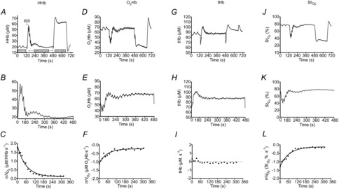
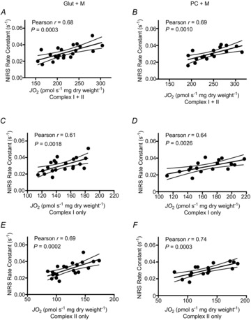
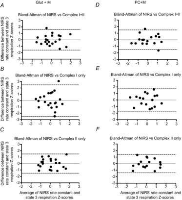
The present study aimed to compare in vivo measurements of skeletal muscle mitochondrial respiratory capacity made using near-infrared spectroscopy (NIRS) with the current gold standard, namely in situ measurements of high-resolution respirometry performed in permeabilized muscle fibres prepared from muscle biopsies. Mitochondrial respiratory capacity was determined in 21 healthy adults in vivo using NIRS to measure the recovery kinetics of muscle oxygen consumption following a ∼15 s isometric contraction of the vastus lateralis muscle. Maximal ADP-stimulated (State 3) respiration was measured in permeabilized muscle fibres using high-resolution respirometry with sequential titrations of saturating concentrations of metabolic substrates. Overall, the in vivo and in situ measurements were strongly correlated (Pearson's r = 0.61-0.74, all P < 0.01). Bland-Altman plots also showed good agreement with no indication of bias. The results indicate that in vivo NIRS corresponds well with the current gold standard, in situ high-resolution respirometry, for assessing mitochondrial respiratory capacity.


Figures

Figure 1
Figure 1
Representative NIRS data NIRS deoxygenated haemoglobin/myoglobin (HHb) signal collected from the vastus lateralis muscle. A, the test protocol consisted of two measurements of resting mVO2, by way of arterial occlusion, post-exercise recovery kinetics of mVO2 and an ischaemic calibration. B, post-exercise recovery kinetics of mVO2 was measured using transient repeated arterial occlusions. C, post-exercise measurements of mVO2 were fit to a monoexponential curve to characterize the rate constant (index of mitochondrial respiratory capacity). Representative NIRS data are also shown for oxygenated haemoglobin/myoglobin (O2Hb), total haemoglobin (tHb) and tissue saturation (StO2) signals for the entire test protocol (D, G and J, respectively), the post-exercise recovery kinetics (E, H and K, respectively) and post-exercise mVO2 measurements (F, I and L, respectively).
Figure 2
Figure 2
Comparison of in vivo (NIRS) and in situ (high-resolution respirometry) indices of skeletal muscle mitochondrial respiratory capacity Correlations between NIRS rate constants for the post-exercise recovery of mVO2 and maximal ADP-stimulated (State 3) respiration in permeabilized muscle fibres from the vastus lateralis muscle in healthy adults in the presence of substrates providing electron flow through Complex I+II (A, glutamate+malate+succinate; D, palmitoyl-l-carnitine+malate+glutamate+succinate), Complex I only (B, glutamate+malate; E, palmitoyl-l-carnitine+malate+glutamate) and Complex II only (C, glutamate+malate+succinate+rotenone; F, palmitoyl-l-carnitine+malate+glutamate+succinate+rotenone); n = 21 for AC; n = 17 for DF. Dashed lines represent 95% confidence intervals.
Figure 3
Figure 3
Level of agreement between in vivo (NIRS) and in situ (high-resolution respirometry) indices of skeletal muscle mitochondrial respiratory capacity Bland–Altman plots (difference vs. average) of in vivo and in situ measurements of skeletal muscle mitochondrial respiratory capacity from the vastus lateralis muscle in healthy adults. Because the two measurement methods have differing units of value, we calculated standardized Z-scores for the study sample. Complex I+II (A, glutamate+malate+succinate; D, palmitoyl-l-carnitine+malate+glutamate+succinate), Complex I only (B, glutamate+malate; E, palmitoyl-l-carnitine+malate+glutamate) and Complex II only (C, glutamate+malate+succinate+rotenone; F, palmitoyl-l-carnitine+malate+glutamate+succinate+rotenone); n = 21 for AC; n = 17 for DF. Dashed lines represent the 95% limits of agreement (mean ± 1.96SD), for which 95% of measurements are expected to lie between.
Figure 4
Figure 4
Simulated data in which the starting, end-exercise, value (y-intercept) is altered A, monoexponential recovery kinetics are such that the initial value has no influence on the change over time (i.e. time and rate constants are the same despite an ∼2-fold difference between starting values). B, the natural log of mVO2 in A plotted against time, in which the slope of the line is equal to the rate constant of the exponential function. Clearly, the two simulated data sets produce equal slopes (parallel lines).

Similar articles

Cited by

References

    1. Belardinelli R, Barstow TJ, Nguyen P, Wasserman K. Skeletal muscle oxygenation and oxygen uptake kinetics following constant work rate exercise in chronic congestive heart failure. Am J Cardiol. 1997;80:1319–1324. - PubMed
    1. Binzoni T, Ferretti G, Schenker K, Cerretelli P. Phosphocreatine hydrolysis by 31P-NMR at the onset of constant-load exercise in humans. J Appl Physiol (1985) 1992;73:1644–1649. - PubMed
    1. Boone J, Koppo K, Barstow TJ, Bouckaert J. Pattern of deoxy[Hb+Mb] during ramp cycle exercise: influence of aerobic fitness status. Eur J Appl Physiol. 2009;105:851–859. - PubMed
    1. Boushel R, Langberg H, Green S, Skovgaard D, Bulow J, Kjaer M. Blood flow and oxygenation in peritendinous tissue and calf muscle during dynamic exercise in humans. J Physiol. 2000;524:305–313. - PMC - PubMed
    1. Brand MD, Nicholls DG. Assessing mitochondrial dysfunction in cells. Biochem J. 2011;435:297–312. - PMC - PubMed

Publication types

LinkOut - more resources