

Widespread grey matter pathology dominates the longitudinal cerebral MRI and clinical landscape of amyotrophic lateral sclerosis
- PMID: 24951638
- PMCID: PMC4132644
- DOI: 10.1093/brain/awu162
Widespread grey matter pathology dominates the longitudinal cerebral MRI and clinical landscape of amyotrophic lateral sclerosis
Abstract
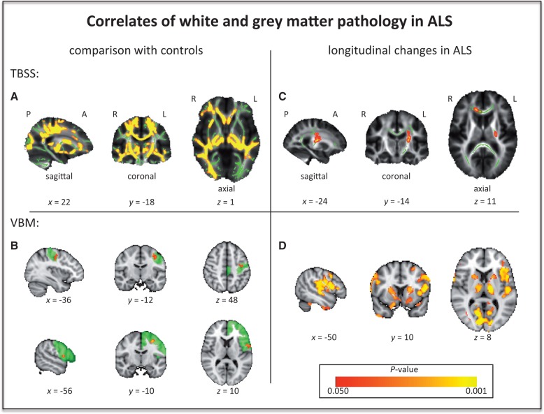
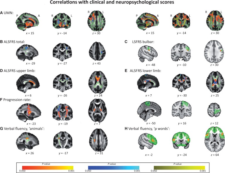
Diagnosis, stratification and monitoring of disease progression in amyotrophic lateral sclerosis currently rely on clinical history and examination. The phenotypic heterogeneity of amyotrophic lateral sclerosis, including extramotor cognitive impairments is now well recognized. Candidate biomarkers have shown variable sensitivity and specificity, and studies have been mainly undertaken only cross-sectionally. Sixty patients with sporadic amyotrophic lateral sclerosis (without a family history of amyotrophic lateral sclerosis or dementia) underwent baseline multimodal magnetic resonance imaging at 3 T. Grey matter pathology was identified through analysis of T1-weighted images using voxel-based morphometry. White matter pathology was assessed using tract-based spatial statistics analysis of indices derived from diffusion tensor imaging. Cross-sectional analyses included group comparison with a group of healthy controls (n = 36) and correlations with clinical features, including regional disability, clinical upper motor neuron signs and cognitive impairment. Patients were offered 6-monthly follow-up MRI, and the last available scan was used for a separate longitudinal analysis (n = 27). In cross-sectional study, the core signature of white matter pathology was confirmed within the corticospinal tract and callosal body, and linked strongly to clinical upper motor neuron burden, but also to limb disability subscore and progression rate. Localized grey matter abnormalities were detected in a topographically appropriate region of the left motor cortex in relation to bulbar disability, and in Broca's area and its homologue in relation to verbal fluency. Longitudinal analysis revealed progressive and widespread changes in the grey matter, notably including the basal ganglia. In contrast there was limited white matter pathology progression, in keeping with a previously unrecognized limited change in individual clinical upper motor neuron scores, despite advancing disability. Although a consistent core white matter pathology was found cross-sectionally, grey matter pathology was dominant longitudinally, and included progression in clinically silent areas such as the basal ganglia, believed to reflect their wider cortical connectivity. Such changes were significant across a range of apparently sporadic patients rather than being a genotype-specific effect. It is also suggested that the upper motor neuron lesion in amyotrophic lateral sclerosis may be relatively constant during the established symptomatic period. These findings have implications for the development of effective diagnostic versus therapeutic monitoring magnetic resonance imaging biomarkers. Amyotrophic lateral sclerosis may be characterized initially by a predominantly white matter tract pathological signature, evolving as a widespread cortical network degeneration.
Keywords: biomarker; diffusion tensor imaging; magnetic resonance imaging; motor neuron disease; voxel-based morphometry.
© The Author (2014). Published by Oxford University Press on behalf of the Guarantors of Brain.
Figures
Comment in
-
Motor neuron disease: Improved imaging biomarkers in amyotrophic lateral sclerosis.Nat Rev Neurol. 2014 Aug;10(8):427. doi: 10.1038/nrneurol.2014.125. Epub 2014 Jul 15. Nat Rev Neurol. 2014. PMID: 25023341 No abstract available.
Similar articles
-
Integration of structural and functional magnetic resonance imaging in amyotrophic lateral sclerosis.Brain. 2011 Dec;134(Pt 12):3470-9. doi: 10.1093/brain/awr279. Epub 2011 Nov 10. Brain. 2011. PMID: 22075069
-
The two-year progression of structural and functional cerebral MRI in amyotrophic lateral sclerosis.Neuroimage Clin. 2017 Dec 18;17:953-961. doi: 10.1016/j.nicl.2017.12.025. eCollection 2018. Neuroimage Clin. 2017. PMID: 29321969 Free PMC article.
-
White matter alterations differ in primary lateral sclerosis and amyotrophic lateral sclerosis.Brain. 2011 Sep;134(Pt 9):2642-55. doi: 10.1093/brain/awr178. Epub 2011 Jul 28. Brain. 2011. PMID: 21798965 Free PMC article.
-
Assessment of the upper motor neuron in amyotrophic lateral sclerosis.Clin Neurophysiol. 2016 Jul;127(7):2643-60. doi: 10.1016/j.clinph.2016.04.025. Epub 2016 May 5. Clin Neurophysiol. 2016. PMID: 27291884 Review.
-
What does imaging reveal about the pathology of amyotrophic lateral sclerosis?Curr Neurol Neurosci Rep. 2015 Jul;15(7):45. doi: 10.1007/s11910-015-0569-6. Curr Neurol Neurosci Rep. 2015. PMID: 26008817 Free PMC article. Review.
Cited by
-
Beyond the Motor Cortex: Thalamic Iron Deposition Accounts for Disease Severity in Amyotrophic Lateral Sclerosis.Front Neurol. 2022 Feb 24;13:791300. doi: 10.3389/fneur.2022.791300. eCollection 2022. Front Neurol. 2022. PMID: 35280261 Free PMC article.
-
New developments and future opportunities in biomarkers for amyotrophic lateral sclerosis.Transl Neurodegener. 2015 Sep 30;4:17. doi: 10.1186/s40035-015-0040-2. eCollection 2015. Transl Neurodegener. 2015. PMID: 26425343 Free PMC article. Review.
-
T1-weighted MRI texture analysis in amyotrophic lateral sclerosis patients stratified by the D50 progression model.Brain Commun. 2024 Nov 5;6(6):fcae389. doi: 10.1093/braincomms/fcae389. eCollection 2024. Brain Commun. 2024. PMID: 39544700 Free PMC article.
-
Limbic Network and Papez Circuit Involvement in ALS: Imaging and Clinical Profiles in GGGGCC Hexanucleotide Carriers in C9orf72 and C9orf72-Negative Patients.Biology (Basel). 2024 Jul 6;13(7):504. doi: 10.3390/biology13070504. Biology (Basel). 2024. PMID: 39056697 Free PMC article.
-
A rare CACNA1H variant associated with amyotrophic lateral sclerosis causes complete loss of Cav3.2 T-type channel activity.Mol Brain. 2020 Mar 6;13(1):33. doi: 10.1186/s13041-020-00577-6. Mol Brain. 2020. PMID: 32143681 Free PMC article.
References
-
- Abrahams S, Goldstein LH, Kew JJ, Brooks DJ, Lloyd CM, Frith CD, et al. Frontal lobe dysfunction in amyotrophic lateral sclerosis. A PET study. Brain. 1996;119(Pt 6):2105–20. - PubMed
-
- Abrahams S, Leigh PN, Harvey A, Vythelingum GN, Grise D, Goldstein LH. Verbal fluency and executive dysfunction in amyotrophic lateral sclerosis (ALS) Neuropsychologia. 2000;38:734–47. - PubMed
-
- Agosta F, Gorno-Tempini ML, Pagani E, Sala S, Caputo D, Perini M, et al. Longitudinal assessment of grey matter contraction in amyotrophic lateral sclerosis: a tensor based morphometry study. Amyotroph Lateral Scler. 2009;10:168–74. - PubMed
-
- Agosta F, Pagani E, Petrolini M, Sormani MP, Caputo D, Perini M, et al. MRI predictors of long-term evolution in amyotrophic lateral sclerosis. Eur J Neurosci. 2010;32:1490–6. - PubMed
Publication types
MeSH terms
Supplementary concepts
Grants and funding
- MR/K006673/1/MRC_/Medical Research Council/United Kingdom
- TURNER/JAN13/944-795/MNDA_/Motor Neurone Disease Association/United Kingdom
- G0701923/MRC_/Medical Research Council/United Kingdom
- MR/K000780/1/MRC_/Medical Research Council/United Kingdom
- MR/K01014X/1/MRC_/Medical Research Council/United Kingdom
LinkOut - more resources
Full Text Sources
Other Literature Sources
Medical
