. 2014 Aug;137(Pt 8):2178-92.
doi: 10.1093/brain/awu142. Epub 2014 Jun 20.

Glycine receptor antibodies in PERM and related syndromes: characteristics, clinical features and outcomes

Affiliations

Glycine receptor antibodies in PERM and related syndromes: characteristics, clinical features and outcomes

Alexander Carvajal-González et al. Brain. 2014 Aug.

Erratum in

  • Brain. 2014 Dec;137(Pt 12):e315. Press, Raomand [corrected to Press, Rayomand]
  • Editorial.
    Kullmann D. Kullmann D. Brain. 2014 Jan;137(Pt 1):1. doi: 10.1093/brain/awt349. Epub 2013 Dec 25. Brain. 2014. PMID: 24371216 Free PMC article. No abstract available.

Abstract

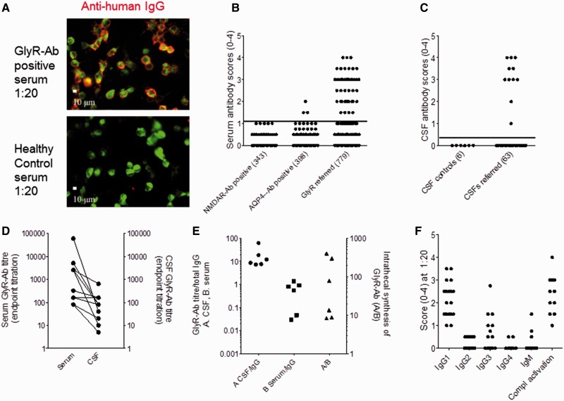
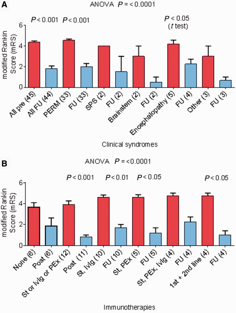
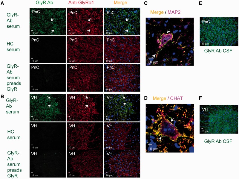
The clinical associations of glycine receptor antibodies have not yet been described fully. We identified prospectively 52 antibody-positive patients and collated their clinical features, investigations and immunotherapy responses. Serum glycine receptor antibody endpoint titres ranged from 1:20 to 1:60 000. In 11 paired samples, serum levels were higher than (n = 10) or equal to (n = 1) cerebrospinal fluid levels; there was intrathecal synthesis of glycine receptor antibodies in each of the six pairs available for detailed study. Four patients also had high glutamic acid decarboxylase antibodies (>1000 U/ml), and one had high voltage-gated potassium channel-complex antibody (2442 pM). Seven patients with very low titres (<1:50) and unknown or alternative diagnoses were excluded from further study. Three of the remaining 45 patients had newly-identified thymomas and one had a lymphoma. Thirty-three patients were classified as progressive encephalomyelitis with rigidity and myoclonus, and two as stiff person syndrome; five had a limbic encephalitis or epileptic encephalopathy, two had brainstem features mainly, two had demyelinating optic neuropathies and one had an unclear diagnosis. Four patients (9%) died during the acute disease, but most showed marked improvement with immunotherapies. At most recent follow-up, (2-7 years, median 3 years, since first antibody detection), the median modified Rankin scale scores (excluding the four deaths) decreased from 5 at maximal severity to 1 (P < 0.0001), but relapses have occurred in five patients and a proportion are on reducing steroids or other maintenance immunotherapies as well as symptomatic treatments. The glycine receptor antibodies activated complement on glycine receptor-transfected human embryonic kidney cells at room temperature, and caused internalization and lysosomal degradation of the glycine receptors at 37°C. Immunoglobulin G antibodies bound to rodent spinal cord and brainstem co-localizing with monoclonal antibodies to glycine receptor-α1. Ten glycine receptor antibody positive samples were also identified in a retrospective cohort of 56 patients with stiff person syndrome and related syndromes. Glycine receptor antibodies are strongly associated with spinal and brainstem disorders, and the majority of patients have progressive encephalomyelitis with rigidity and myoclonus. The antibodies demonstrate in vitro evidence of pathogenicity and the patients respond well to immunotherapies, contrasting with earlier studies of this syndrome, which indicated a poor prognosis. The presence of glycine receptor antibodies should help to identify a disease that responds to immunotherapies, but these treatments may need to be sustained, relapses can occur and maintenance immunosuppression may be required.

Keywords: autoantibody; autoimmune encephalitis; glycine receptor; progressive encephalomyelitis with rigidity and myoclonus; stiff person syndrome.


Figures

Figure 1
Figure 1
GlyR antibodies (Ab) in PERM and related disorders. (A) A patient’s serum IgG binding to HEK293 cells expressing GlyRα1-EGFP (green); the IgG binding is detected with anti-human IgG (red). No IgG binding is observed with serum from a healthy individual. (B) Visual scores of sera (diluted 1:20) referred for routine GlyR antibody testing compared with scores in sera positive for NMDAR or aquaporin 4 (AQP4) antibodies studied as neurological controls. (C) Scores of referred CSF samples (diluted 1:1) compared with multiple sclerosis control CSFs. (D) Endpoint titrations for sera and paired CSFs; the CSF titres are generally lower than serum titres but in three patients are equal or only a dilution lower. (E) Calculation of intrathecal synthesis in six paired CSFs. GlyR antibody titres (from D) divided by IgG concentration for sera (serum/IgG) or for CSFs (CSF/IgG). Intrathecal synthesis rates are given as the ratio between these values as described above, and are raised in all patients, particularly in three. (F) Scores for each IgG subtype and for the deposition of complement C3 on the GlyRα1-expressing HEK cells. The antibodies are mainly IgG1 and IgG3 subtypes and activate complement on the surface of the GlyRα1-transfected cells.
Figure 2
Figure 2
Modified Rankin scale (mRS) scores at maximum severity and at recent follow-up in patients with PERM or related syndromes. One patient was lost to long-term follow-up. (A) Modified Rankin scale scores according to the clinical syndrome compared between maximum severity and follow-up in all patients including the four who died (modified Rankin scale 6). (B) Modified Rankin scale scores before and after different immunotherapies for all patients, excluding the four who died. Further details are available in Table 4. FU = follow-up; PEx = plasma exchange; IvIg = intravenous immunoglobulins; St = steroids.
Figure 3
Figure 3
GlyR antibodies and GlyRα1s are internalized in HEK293 cells and GlyRα1 is targeted to the lysosomal pathway. (A) GlyR antibodies (Ab; red) bound to the surface of GlyRα1-EGFP (green) transfected HEK cells (shown at 0 h and 16 h). Loss of surface bound GlyR antibody is seen by 16 h when the cells are incubated at 37°C, but not when incubated at 4°C, or when fixed before incubation. (B) Results from four GlyR antibody-positive sera compared with four healthy individuals’ (HC) sera with the surface IgG binding scored after different incubation times. Two-way ANOVA (F = 7.832, df = 20, P < 0.0001). Significant differences are marked with asterisks. (C) By 2 h of incubation at 37°C, the surface bound GlyR antibodies (green) are partly lost but can be detected after fixing and permeabilization (perm) with anti-human IgG (red). Unpermeabilized (Not perm) cells did not show internalized IgG. (D) The percentage of cells with internalized GlyR antibody at 0 and 2 h. Results from two sera were compared between extracellular (green) and intracellular (red) IgG using a two-sided students t-test (*t = 9.448, df = 18, P < 0.0001; **t = 8.707, df = 18, P < 0.0001). (E) GlyR antibody binding causes internalization of the GlyRα1. After 1 h treatment with cyclohexamide, HEK cells expressing GlyRα1-EGFP were incubated at 37°C with patient or healthy control sera for 2 h. Incubation with patient serum led to a decrease in the percentage of cells expressing GlyRα1-EGFP (upper row), but not with control serum (lower row), as shown also at higher resolution on the right in cells post-labelled with DAPI (blue). (F) Quantification of GlyRα1-EGFP internalization from two experiments with a single serum. Groups were compared using a two-sided Student’s t-test (**t = 10.15, df = 18, P < 0.0001; *t = 15.01, df = 18, P < 0.0001). (G) Internalized GlyRα1-EGFP is targeted to the lysosome. The GlyRα1-EGFP expressing HEK cells were labelled with antibodies to the late endosomal marker (LAMP2). Internalized GlyRα1-EGFP (green) co-localized with LAMP2 (red) in cells exposed to patients’ serum but co-localization was much less in cells exposed to healthy control serum. (H) The percentage of cells with co-localization of GlyRα1-EGFP and LAMP2 from two experiments with one serum from GlyR antibody and one healthy control. Groups were compared using a two-sided Students t-test (*t = 8.515, df = 18, P < 0.0001; **t = 13.73, df = 18, P < 0.0001).
Figure 4
Figure 4
GlyR antibody-positive patient serum from a typical PERM patient binds to CNS regions involved in motor regulation. (A) Top row: Double labelling of neurons in the pontine reticular nucleus (PnC) of the brainstem with patient serum (green) and monoclonal antibody to the GlyRα1 (red) show colocalization on the neuronal soma and in the neuropil (arrows); nuclei are stained with DAPI (blue). Middle row: Healthy control serum did not bind to the pontine reticular nucleus. Lower row: After pre-adsorption against HEK-GlyRα1 cells there was a marked decrease in pontine reticular nucleus staining. (B) Similar results are shown for binding to the ventral horn of the spinal cord. (C and D) Confocal images show the patient’s serum (green) colocalizing with GlyRα1 monoclonal antibody (red) on the surface of a large neuron in the pontine reticular nucleus, which is labelled with the cytoplasmic neuronal marker anti-MAP2 antibody (purple), or on a motor neuron in the ventral horn of the spinal cord labelled with motor neuron marker anti-choline acetyltranferase antibody [CHAT (purple)]. (E and F) CSF from the same patient binds in a similar pattern. Results of seven representative patients are summarized in Supplementary Table 2. HC = healthy control.

Comment in

References

    1. Alexopoulos H, Dalakas MC. A critical update on the immunopathogenesis of Stiff Person Syndrome. Eur J Clin Invest. 2010;40:1018–25. - PubMed
    1. Alexopoulos H, Akrivou S, Dalakas MC. Glycine receptor antibodies in stiff-person syndrome and other GAD-positive CNS disorders. Neurology. 2013;81:1962–4. - PubMed
    1. Angus-Leppan H, Rudge P, Mead S, Collinge J, Vincent A. Autoantibodies in sporadic Creutzfeldt-Jakob disease. JAMA Neurol. 2013;70:919–22. - PubMed
    1. Araki T, Yamano M, Murakami T, Wanaka A, Betz H, Tohyama M. Localization of glycine receptors in the rat central nervous system: an immunocytochemical analysis using monoclonal antibody. Neuroscience. 1988;25:613–24. - PubMed
    1. Bourke D, Roxburgh R, Vincent A, Cleland J, Jeffery O, Dugan N, et al. Hypoventilation in glycine-receptor antibody related progressive encephalomyelitis, rigidity and myoclonus. J Clin Neurosci. 2014;21:876–8. - PubMed

Publication types

MeSH terms

Supplementary concepts