

Fluorescence cryo-microscopy: current challenges and prospects
- PMID: 24951858
- PMCID: PMC4094034
- DOI: 10.1016/j.cbpa.2014.05.007
Fluorescence cryo-microscopy: current challenges and prospects
Abstract
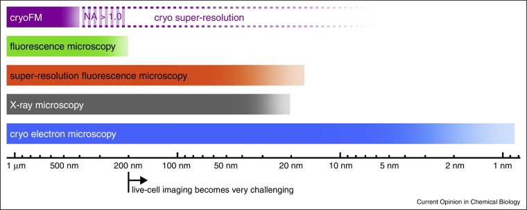
Studying biological structures with fine details does not only require a microscope with high resolution, but also a sample preparation process that preserves the structures in a near-native state. Live-cell imaging is restricted mostly to the field of light microscopy. For studies requiring much higher resolution, fast freezing techniques (vitrification) are successfully used to immobilize the sample in a near-native state for imaging with electron and X-ray cryo-microscopy. Fluorescence cryo-microscopy combines imaging of vitrified samples with the advantages of fluorescence labeling of biological structures. Technical considerations as well as the behavior of fluorophores at low temperatures have to be taken into account for developing or adapting super-resolution methods under cryo conditions to exploit the full potential of this technique.
Copyright © 2014 The Authors. Published by Elsevier Ltd.. All rights reserved.
Figures
References
-
- Erbe E.F., Rango A., Foster J., Josberger E.G., Pooley C., Wergin W.P. Collecting, shipping, storing, and imaging snow crystals and ice grains with low-temperature scanning electron microscopy. Microsc Res Tech. 2003;62:19–32. - PubMed
-
- Diller K.R., Cravalho E.G. A cryomicroscope for the study of freezing and thawing processes in biological cells. Cryobiology. 1970;7:191–199. - PubMed
-
- Holt W.V., Head M.F., North R.D. Freeze-induced membrane damage in ram spermatozoa is manifested after thawing: observations with experimental cryomicroscopy. Biol Reprod. 1992;46:1086–1094. - PubMed
-
- Moerner W.E., Orrit M. Illuminating single molecules in condensed matter. Science. 1999;283:1670–1676. - PubMed
-
- Kozankiewicz B., Orrit M. Single-molecule photophysics, from cryogenic to ambient conditions. Chem Soc Rev. 2014;43:1029–1043. - PubMed
-
This review discusses the photophysics of fluorescent molecules at low and ambient temperatures at the single molecule level.
Publication types
MeSH terms
Substances
Grants and funding
LinkOut - more resources
Full Text Sources
Other Literature Sources
