. 2014 Jul;16(7):663-72.
doi: 10.1038/ncb2988. Epub 2014 Jun 22.

The kinesin-4 protein Kif7 regulates mammalian Hedgehog signalling by organizing the cilium tip compartment

Affiliations

The kinesin-4 protein Kif7 regulates mammalian Hedgehog signalling by organizing the cilium tip compartment

Mu He et al. Nat Cell Biol. 2014 Jul.

Abstract

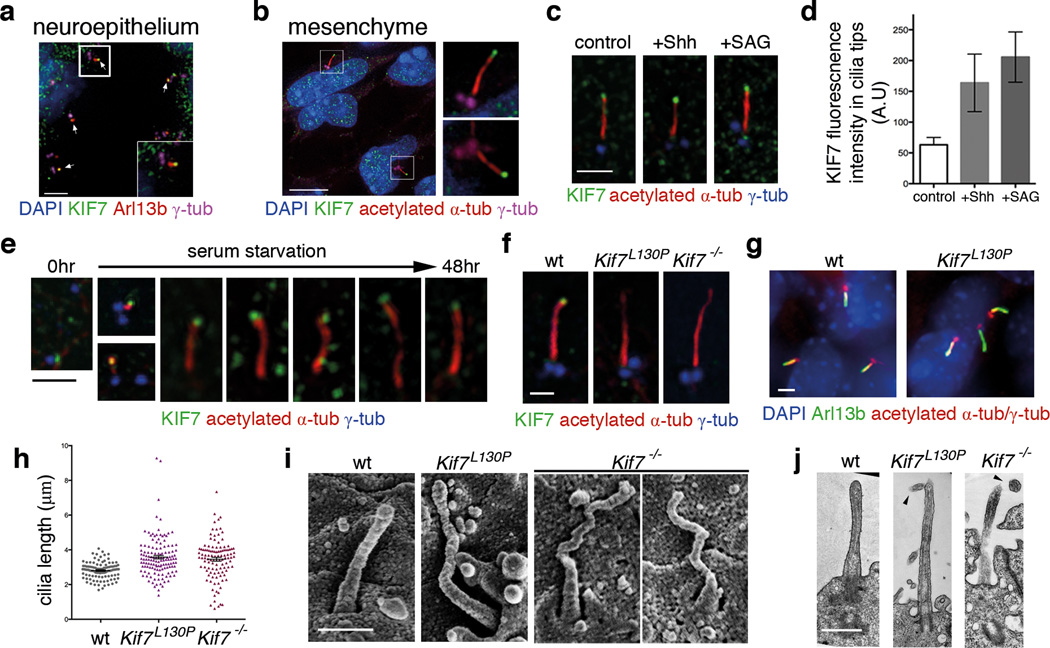
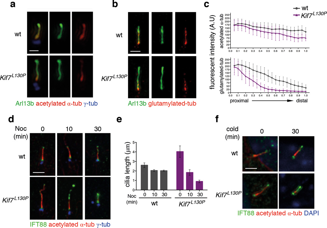
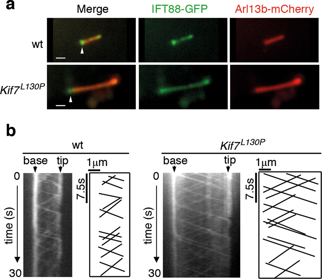
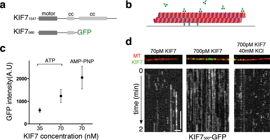
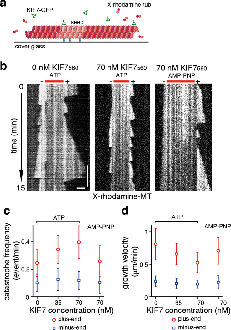
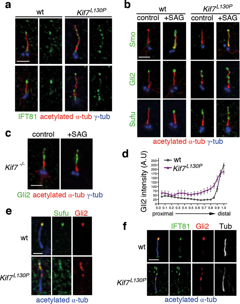
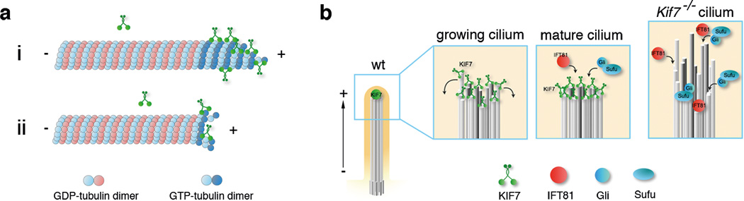
Mammalian Hedgehog (Hh) signal transduction requires a primary cilium, a microtubule-based organelle, and the Gli-Sufu complexes that mediate Hh signalling, which are enriched at cilia tips. Kif7, a kinesin-4 family protein, is a conserved regulator of the Hh signalling pathway and a human ciliopathy protein. Here we show that Kif7 localizes to the cilium tip, the site of microtubule plus ends, where it limits cilium length and controls cilium structure. Purified recombinant Kif7 binds the plus ends of growing microtubules in vitro, where it reduces the rate of microtubule growth and increases the frequency of microtubule catastrophe. Kif7 is not required for normal intraflagellar transport or for trafficking of Hh pathway proteins into cilia. Instead, a central function of Kif7 in the mammalian Hh pathway is to control cilium architecture and to create a single cilium tip compartment, where Gli-Sufu activity can be correctly regulated.


Figures

Figure 1
Figure 1. KIF7 constrains the length of primary cilia
(a–b) KIF7 (green) localizes to the tips of primary cilia in e10.5 wild-type embryos. Arl13b (red) marks cilia in (a) and acetylated-α-tubulin (red) marks microtubule axoneme in (b). γ-tubulin (magenta) marks basal bodies. DAPI (blue) labels nuclei. Scale bar = 5µm. (c) In wild-type MEFs, KIF7 (green) is enriched in tips of primary cilia upon treatment with Shh-N recombinant protein (2µg/ml) or SAG (100nM) for 24 hours. Scale bar = 2µm. (d) Quantitation of KIF7 fluorescence intensity shown in (C). n = 150 cilia pooled from 3 independent experiments were counted for each condition. The error bars represent the SD. p<0.0001 between control and SAG or Shh treatments by one-way ANOVA. (e) KIF7 (green) is associated with one of the two centrioles prior to ciliogenesis and localizes to the tips of primary cilia throughout cilia elongation induced by serum starvation in wild-type MEFs. (f) KIF7 is not detected at the tips of Kif7L130P or Kif7−/− MEF cilia. Acetylated-α-tubulin (red) marks primary cilia and γ-tubulin (blue) marks basal bodies in (c), (e) and (f). Scale bar = 1µm in (c), (e) and (f). (g) Primary cilia of wild-type and Kif7L130P MEFs stained with Arl13b (green), acetylated-α-tubulin and γ-tubulin (red). DAPI in blue. Scale bar = 2µm. (h) Measurements of MEF cilia length between wild-type and Kif7 mutants using Arl13b as cilia marker. >100 cilia pooled from 4 independent experiments were counted for each genotype (n=108 cilia for wild type, n=133 cilia for Kif7L130P mutant, and n=111 for Kif7−/− mutant). p<0.0001 between wild-type cilia and Kif7 mutant cilia by one-way ANOVA. The error bars represent the SEM. (i) Scanning electron microscopy shows neural tube cilia of e10.5 wild-type, Kif7L130P and Kif7−/− embryos. Wild-type neural cilia were 0.97±0.17 µm and Kif7L130P neural cilia were approximately 1.2±0.28µm long (n=50 for each genotype). Scale bar = 0.5µm. (j) TEM images of longitudinal sections of wild-type, Kif7L130P and Kif7−/− neural tube cilia. Arrowheads point to twisted tips of mutant cilia. Scale bar = 0.5µm.
Figure 2
Figure 2. KIF7 stabilizes primary cilia
(a) Tubulin acetylation (red) and (b) glutamylation (red) are reduced at the distal segment of cilia in Kif7L130P MEFs compared to wild type. Arl13b (green) labels the entire cilia in (a–b) and γ-tubulin (blue) marks basal bodies in (a). Scale bar = 1µm. (b) Fluorescence intensities of acetylation and glutamylation, as shown in panel (a–b), normalized for cilia length. n= 50 cilia were measured for each genotype pooled from 3 independent experiments. The error bars represent the SD. For tubulin acetylation, p<0.001 for the distal half of the cilia. For tubulin glutamylation, p<0.0001 for the distal 80% of the cilia. (d) Time course of cilia shortening in response to nocodazole treatment in wild-type and Kif7L130P MEFs. IFT88 (green) accumulated distally Kif7L130P MEF cilia during cilia retraction. γ-tubulin (blue) marks basal bodies. Scale bar = 2µm. (e) Cilia length was measured using acetylated-α-tubulin from the time-course experiment shown in (d). n=30 for each condition and each time point pooled from 3 independent experiments. The error bars represent the SD ( p<0.001 between wild-type and Kif7L130P at 0 and 30 minutes; n.s between two genotypes at 10 minutes). (f) Time course of cilia retraction at 4°C in wild-type and Kif7L130P MEFs. Cilia are marked with acetylated-α-tubulin (red) and IFT88 (green). DAPI in blue. Scale bar = 2µm.
Figure 3
Figure 3. KIF7 does not affect the rates of intraflagellar transport (IFT)
(a) Still images of wild-type and Kif7L130P primary cilium expressing IFT88-GFP (green) and Arl13b-mCherry (red) before live imaging. Arrowheads mark the base of the ciliary axoneme. Scale bar = 1µm. (b) Kymographs generated from time lapse imaging of wild-type and Kif7L130P primary cilia. Also see Supplementary Movie. S1 and S2. The base and tip of the cilium are indicated with arrowheads. Horizontal scale bar (distance) = 1µm. Vertical scale bar (time) = 7.5 seconds.
Figure 4
Figure 4. A purified KIF7 motor construct lacks detectable microtubule-dependent motility
(a) Domain organizations of full-length KIF7 and the N-terminal fragment used for in vitro assays. (b) Schematic of the assay for examining KIF7-microtubule interaction. X-rhodamine labeled microtubules (red) were attached to a PEG-passivated glass surface by biotin–NeutrAvidin (gray and blue) links prior to addition of KIF7 motor protein (green). (c) Average fluorescent intensity of microtubule-bound KIF7560-GFP plotted against KIF7 protein concentration in the presence of either 1 mM MgATP or 2 mM AMP-PNP. n = 20 microtubules for each condition were analyzed. Error bars represent SD. p<0.0001 by one-way ANOVA. (d) KIF7560-GFP gliding assay (70 pM or 700 pM) under different ionic strength conditions. Representative kymographs from assays at BRB80 and BRB80 +40 mM KCl buffer conditions in the presence of 1 mM MgATP. Still images show KIF7560-GFP (green) binding to X-rhodamine labeled microtubules (red). Kymographs of KIF7560-GFP are shown in gray scale. Horizontal scale bar = 2µm. Vertical scale bar = 30s. The frame rate was 1 frame per 0.5 s.
Figure 5
Figure 5. KIF7 alters microtubule dynamics in vitro
(a) Schematic of the experimental setup of microtubule dynamics assay: X-rhodamine- and biotin-labeled microtubule seeds, which were polymerized in the presence of GMP-CPP, were immobilized on a glass coverslip, and then incubated with a mixture of X-rhodamine labeled tubulin (1.8 µM) and non-fluorescent tubulin (18 µM; 1 mM MgGTP). (b) Representative kymographs of X-rhodamine-labeled microtubules (show in grey scale) polymerizing in the absence or presence of KIF7560-GFP at the indicated concentrations in the presence of either 1 mM MgATP or 2 mM AMP-PNP. The frame rate was 1 frame per 5s. Microtubule seed and polarity are indicated above each kymograph. Horizontal scale bar = 2µm. Vertical scale bar = 180s. (c) The frequencies of microtubule catastrophe in the absence of KIF7 (n=272), 35 nM KIF560-GFP (n=263; p<0.0001 compared to control), 70 nM KIF560-GFP (n=492; p<0.0001 compared to control and p<0.01 compared to 35 nM KIF560-GFP) in the presence of 1 mM MgATP and 70 nM KIF560-GFP (n=179; n.s compared to control) in the presence of 2 mM AMP-PNP were plotted against the KIF7560-GFP concentration. The catastrophe frequencies of microtubule minus-ends were not affected by KIF560-GFP (n=82 for control, n=51 for 35nM KIF560-GFP with Mg ATP, n=94 for 70 nM KIF560-GFP with MgATP and n=53 for 70 nM KIF560-GFP with AMP-PNP were analyzed; n.s compared to control). (d) Microtubule growth in the absence of KIF7 (n=145), 35 nM KIF7560-GFP (n=109; p<0.0001 compared to control), 70 nM (n=263; p<0.0001 compared to control and p < 0.001 compared to 35 nM KIF7560-GFP) in the presence of 1 mM MgATP and 70nM KIF7560-GFP (n=101; p<0.01 compared to control) in the presence of 2 mM AMP-PNP were plotted against the KIF7560-GFP concentration. The growth rates of microtubule minus-ends were not affected by KIF560-GFP (n=51 for control, n=47 for 35nM KIF560-GFP with Mg ATP, n=45 for 70 nM KIF560-GFP with MgATP and n=51 for 70 nM KIF560-GFP with AMP-PNP were analyzed; n.s compared to control). Data sets pooled from 3 independent experiments were analyzed. Error bars represent the SD. p values were calculated by nonparametric test for multiple comparisons.
Figure 6
Figure 6. KIF7 tracks microtubule plus-ends
Representative images showing KIF7560-GFP (70 nM) associated with dynamic microtubules in the presence of (a) 1 mM MgATP or (b) 2 mM AMP-PNP. Arrows indicates microtubules used for line-scan based fluorescence intensity analysis. Scale bar = 2µm. Also see Supplementary Movie S3. (c) Representative kymographs of KIF7560-GFP on X-rhodamine-labeled dynamic microtubules in the presence of 1 mM MgATP (n=263; two examples shown) and (d) 2 mM AMP-PNP (n=101). Plus-ends of microtubules are to the right on the kymographs in (c) and (d). Horizontal scale bar = 2µm. Vertical scale bar = 180s. The frame rate was 1 frame per 5s. Data sets from 2–3 independent experiments were analyzed.
Figure 7
Figure 7. Multiple cilia tip-like compartments form in the absence of KIF7
(a) IFT81 (green) localization in the wild-type and Kif7L130P cilia. (b) Cilia localization of Smo, Gli2 and Sufu in wild-type and Kif7−/− MEFs in response to pathway activation by 100nM SAG treatment for 24 hr. (c) Gli2 localizes to puncta along Kif7−/− ciliary axonemes independent of pathway activation. Acetylated-α-tubulin (red) marks axonemal microtubules and γ-tubulin (red) marks basal bodies in (a) to (c). (d) Distribution of fluorescence intensity of Gli2 in SAG-treated wild-type and Kif7L130P cilia, normalized to cilia length. n=50 cilia were analyzed for each condition pooled from 3 independent experiments. p<0.0001. The error bars represent the SEM. (e) Sufu (green) and Gli2 (red) co-localization in the tip of wild-type cilia. In Kif7L130P mutant cilia, Sufu and Gli2 co-localize to the same puncta along the axoneme. (f) IFT81 (green) and Gli2 (red) co-localization in the tip of wild-type cilia. In Kif7L130P mutant cilia, IFT81 and Gli2 co-localize to the same puncta along the axoneme. Acetylated-α-tubulin shown in blue in merged panels and in gray-scale in separated channels. Scale bars = 1µm in (a)–(c) and (e)–(f).
Figure 8
Figure 8. Model for KIF7-dependent regulation of plus-end dynamics in primary cilia
(a) Proposed model for the regulation of microtubule dynamics by KIF7. i: KIF7 preferentially binds to the GTP-bound tubulin at microtubule plus-ends, inhibiting addition of tubulin dimers and microtubule growth. ii: By destabilizing the GTP-bound tubulin cap at microtubule plus-ends, KIF7 can promote catastrophe and thereby limit microtubule growth. (b) Model for KIF7 function at the tip of primary cilia. In wild type, KIF7 acts at the distal end of the growing cilium to prevent overgrowth of individual microtubules and to coordinate the growth of 9-doublet microtubules; only organized microtubule arrays are a suitable substrate for post-translational modifications. In the mature cilium, KIF7 creates a single distal tip compartment where IFT81 and Gli-Sufu complexes are enriched. In the absence of KIF7, growth of axonemal microtubules is not limited and synchronized, leading to longer and unstable cilia and ectopic tip-like compartments that contain IFT81 along the axoneme. Gli-Sufu complexes localized to the ectopic tip compartments, where they can be inappropriately activated in the absence of Shh ligand.

Comment in

Similar articles

Cited by

References

    1. Bieling P, Telley IA, Surrey T. A minimal midzone protein module controls formation and length of antiparallel microtubule overlaps. Cell. 2010;142:420–432. - PubMed
    1. Hu C-K, Coughlin M, Field CM, Mitchison TJ. KIF4 regulates midzone length during cytokinesis. Curr. Biol. 2011;21:815–824. - PMC - PubMed
    1. Stumpff J, Wagenbach M, Franck A, Asbury CL, Wordeman L. Kif18A and chromokinesins confine centromere movements via microtubule growth suppression and spatial control of kinetochore tension. Dev. Cell. 2012;22:1017–1029. - PMC - PubMed
    1. Subramanian R, Ti S-C, Tan L, Darst SA, Kapoor TM. Marking and measuring single microtubules by PRC1 and kinesin-4. Cell. 2013;154:377–390. - PMC - PubMed
    1. van der Vaart B, et al. CFEOM1-Associated Kinesin KIF21A Is a Cortical Microtubule Growth Inhibitor. Dev. Cell. 2013 - PubMed

Publication types

MeSH terms