. 2014 Aug;28(8):841-50.
doi: 10.1007/s10822-014-9765-8. Epub 2014 Jun 22.

Design of a colicin E7 based chimeric zinc-finger nuclease

Affiliations

Design of a colicin E7 based chimeric zinc-finger nuclease

Eszter Németh et al. J Comput Aided Mol Des. 2014 Aug.

Abstract

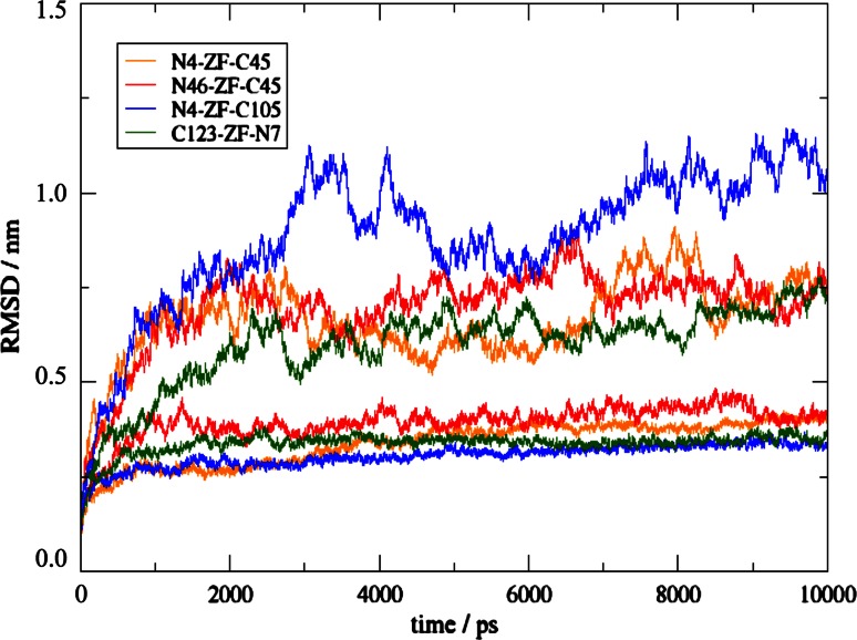
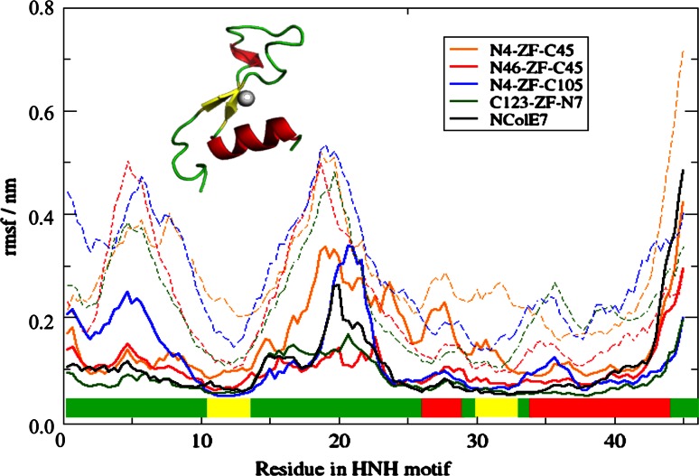
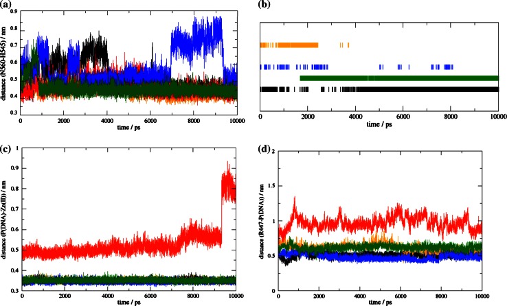
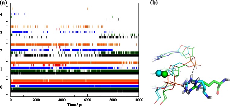
Colicin E7 is a natural bacterial toxin. Its nuclease domain (NColE7) enters the target cell and kills it by digesting the nucleic acids. The HNH-motif as the catalytic centre of NColE7 at the C-terminus requires the positively charged N-terminal loop for the nuclease activity-offering opportunities for allosteric control in a NColE7-based artificial nuclease. Accordingly, four novel zinc finger nucleases were designed by computational methods exploiting the special structural features of NColE7. The constructed models were subjected to MD simulations. The comparison of structural stability and functional aspects showed that these models may function as safely controlled artificial nucleases. This study was complemented by random mutagenesis experiments identifying potentially important residues for NColE7 function outside the catalytic region.


Figures

Fig. 1
Fig. 1
Two possible complexes for the design of a ZFN, in “straight” (a) and “reverse” (b) orientations. NColE7 in green and ZF protein consisting of three fingers in orange. The N- and C-termini of the proteins are marked in blue and red, respectively. The proposed links at positions S1, S2, R1 and R2 are indicated in Tables S2 and S3
Fig. 2
Fig. 2
Sequences of the designed ZFNs. The ZF proteins are in blue, while NColE7 is divided into three parts: the original N-terminus (in red), the middle part not used in the model (in grey, crossed out) and the original C-terminal part (in green). The linkers are shown with black bold letters. The HNH motif of NColE7 is underlined. The N4–ZF–C105, N4–ZF–C45 and N46–ZF–C45 models are in the reverse orientation, while C123–ZF–N7 is the straight joint of protein sequences. For explanation of the names see the text
Fig. 3
Fig. 3
Location of the random mutations, as shown in the crystal structure 3FBD (D493Q–NColE7 in complex with a 18 bp DNA). The residues in red occur as single site mutations, while the orange ones are double site mutations
Fig. 4
Fig. 4
Designed ZFN structures, as starting points of MD simulations. The N-terminal part of NColE7 is shown in red, the C-terminal part in green, the ZF-s in blue, and the linkers are shown in black. The grey spheres indicate Zn2+-ions. a N4–ZF–C45 (reverse), b N46–ZF–C45 (reverse), c N4–ZF–C105 (reverse), d C123–ZF–N7 (straight). The C- and N-termini of the ZFNs are marked by “C” and “N”, respectively
Fig. 5
Fig. 5
Atom-positional RMSD of the ZFN models in the simulations in complex with DNA (lower curve in each color) and in the control simulations of the proteins without the DNA (higher curves in each color)
Fig. 6
Fig. 6
Atom-positional RMSFs of residues in the HNH motif of ZFN-s and NColE7 as a reference. The residue numbers are shifted to 1–45 in all models for comparison, corresponding to the HNH residues 532–576 in the NColE7 numbering. Dashed lines indicate the control simulations for proteins without DNA. The secondary structural elements of HNH motif are marked above the x axis, showing the loops in green, α-helices in red and β-sheets in yellow
Fig. 7
Fig. 7
a Distance of residues corresponding to N560 (CG) and H545 (CA) in NColE7 as a function of time. b Presence of H-bond between the residues corresponding to H545 and V555 in NColE7. c Catalytic distances as a function of time: Zn2+-O (at the scissile phosphate) and d R447(NE)-P (scissile phosphate). In all panels, NColE7 is represented by black curves, N4–ZF–C45 by orange, N46–ZF–C45 by red, N4–ZF–C105 by blue and C123–ZF–N7 by green
Fig. 8
Fig. 8
a The number of H-bonds between the arginine corresponding to R447 in NColE7 and the DNA atoms in the different models as a function of time. Numbers on the y axis indicate how many hydrogen bonds were found at a given time. NColE7 is in black, N4–ZF–C45 in orange, N46–ZF–C45 in red, N4–ZF–C105 in blue and C123–ZF–N7 in green. b The interactions of the controlling Arg at 2,000 ps (in green) and 8,500 ps (in blue) in the N4–ZF–C45 simulation

Similar articles

Cited by

References

    1. Kim YG, Cha J, Chandrasegaran S. Hybrid restriction enzymes: zinc finger fusions to Fok I cleavage domain. Proc Natl Acad Sci USA. 1996;93:1150–1160. - PMC - PubMed
    1. Ramalingam S, Kandavelou K, Rajenderan R, Chandrasegaran S. Creating designed zinc-finger nucleases with minimal cytotoxicity. J Mol Biol. 2011;405:630–641. doi: 10.1016/j.jmb.2010.10.043. - DOI - PMC - PubMed
    1. Moehle EA, Rock JM, Lee YL, Jouvenot Y, DeKelver RC, Gregory PD, Urnov FD, Holmes MC. Targeted gene addition into a specified location in the human genome using designed zinc finger nucleases. Proc Natl Acad Sci USA. 2007;104:3055–3060. doi: 10.1073/pnas.0611478104. - DOI - PMC - PubMed
    1. Sharan SK, Thomason LC, Kuznetsov SG, Court DL. Recombineering: a homologous recombination-based method of genetic engineering. Nat Protoc. 2009;4:206–223. doi: 10.1038/nprot.2008.227. - DOI - PMC - PubMed
    1. Connelly JP, Barker JC, Pruett-Miller S, Porteus MH. Gene correction by homologous recombination with zinc finger nucleases in primary cells from a mouse model of a generic recessive genetic disease. Mol Ther. 2010;18:1103–1110. doi: 10.1038/mt.2010.57. - DOI - PMC - PubMed

Publication types

LinkOut - more resources