. 2014 Aug;10(8):686-91.
doi: 10.1038/nchembio.1561. Epub 2014 Jun 22.

A G-quadruplex-containing RNA activates fluorescence in a GFP-like fluorophore

Affiliations

A G-quadruplex-containing RNA activates fluorescence in a GFP-like fluorophore

Hao Huang et al. Nat Chem Biol. 2014 Aug.

Abstract

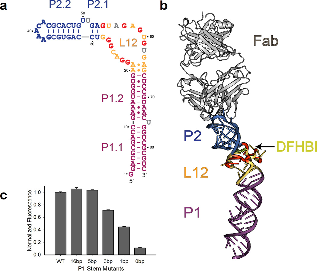
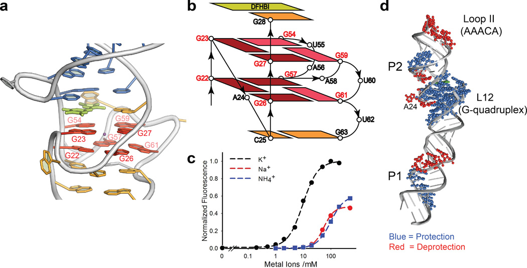
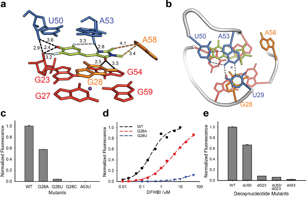
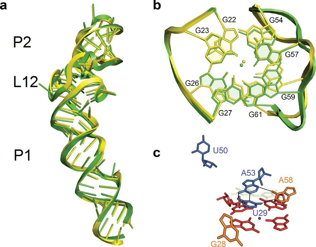
Spinach is an in vitro-selected RNA aptamer that binds a GFP-like ligand and activates its green fluorescence. Spinach is thus an RNA analog of GFP and has potentially widespread applications for in vivo labeling and imaging. We used antibody-assisted crystallography to determine the structures of Spinach both with and without bound fluorophore at 2.2-Å and 2.4-Å resolution, respectively. Spinach RNA has an elongated structure containing two helical domains separated by an internal bulge that folds into a G-quadruplex motif of unusual topology. The G-quadruplex motif and adjacent nucleotides comprise a partially preformed binding site for the fluorophore. The fluorophore binds in a planar conformation and makes extensive aromatic stacking and hydrogen bond interactions with the RNA. Our findings provide a foundation for structure-based engineering of new fluorophore-binding RNA aptamers.


Figures

Figure 1
Figure 1. Global structure of the Spinach RNA-Fab complex
(a). Observed secondary structure of Spinach construct containing G37AAACAC43 antigenic tag (bold blue letters). The L12 region (brown-yellow) contains a G-quadruplex motif, with participating Gs in bold red letters. Flipped-out nucleotides with partial electron densities are in grey. (b). Overview of the Spinach RNA structure in complex with the BL3-6 Fab (grey). The RNA forms a long, slightly bent helical domain that docks into the Fab heavy chain CDRs via binding interactions with the GAAACAC tag (blue). The core G-quadruplex region in L12, colored yellow and red, forms a platform for stacking of the DHFBI ligand (lemon). (c). Fluorescence activation by P1 stem truncation mutants.Data represent mean values ± s.d. from three measurements. The entire P1 stem (P1.1 and P1.2) is replaced with a designated number of Watson-Crick base pairs in each truncate as shown in Supplementary Fig. 10. A Spinach construct containing a five base-pair P1 stem retains WT levels of fluorescence activation. Sequences of them and other mutants are all included in Supplementary Table 3.
Figure 2
Figure 2. The Two-Layer G-quadruplex in L12 of Spinach
(a). Highlight of the G-quadruplex (red); The G-quadruplex forms a platform for stacking of the DFHBI fluorophore (lemon). A potassium ion (purple) sits in the center of the quadruplex between two layers. The color codes of other nucleotides match Fig. 1a. (b). Topological diagram of the two-layer G-quadruplex region. The anti-glycosidic guanines are highlighted with dark red. (c). Monovalent cationdependence of fluorescence activation. A fit of the data to the Hill equation gave an apparent midpoint (K1/2 = 9.6 ± 0.4 mM) and Hill slope (n) of 1.6 ± 0.1 for potassium. (d). SHAPE analysis outcome of Spinach.The SHAPE footprints of the standard-folded WT Spinach [Mg+/K+/DFH+] (Supplementary Fig. 12 for the original gel image) are superimposed onto the crystal structure. A more detailed interpretation of this data is in Online Methods.
Figure 3
Figure 3. The fluorophore binding site in Spinach
(a). Side view of the DFHBI binding site. U29 is omitted for clarity. Black dashes (with the distances numbered in Å) represent inferred hydrogen bonds from DFHBI to the G28 nucleobase and several 2’-hydroxyl groups. Potential hydrogen bonds from fluorine are in grey dashes with distances. The imidazolinone methyl groups of DFHBI may engage in hydrophobic interactions (orange dashes) with A58 (orange). Ligand atoms: C = lemon, N = blue, F = cyan, O = red. DFHBI geometry, positioning, and orientation were deduced as described in Online Methods and Supplementary Figs. 13, 14, and 15, respectively. (b). Top-down view showing DFHBI (lemon) and adjacent nucleotide G28 stacking between a U50-A53-U29 Hoogsteen base triple (blue) above and a layer of G-quadruplex (red) below. (c). Mutations at G28 or A53 diminish fluorescence activation. Data represent mean values ± s.d. from three measurements. DFHBI concentration was 5 µM. (d). Dependence of fluorescence on DFHBI concentration. Fits of the data to the Hill equation give KD = 300 ± 68 nM and 4.4 ± 0.8 µM for WT and G28A, respectively. Weak fluorescence activation by the G28U mutant precluded KD determination. (e). Effect of deoxynucleotide mutants on corresponding positions (G23, U50 and A53) in Spinach. Data represent mean values ± s.d. from three measurements.
Figure 4
Figure 4. The structure of Spinach RNA in the absence of DFHBI
(a). Overlay of Spinach structures obtained in the presence (green) and absence (yellow) of bound DFHBI fluorophore. DFHBI binding has minimal influence on global architecture (RMS = 0.92Å on RNA). (b). Overlay of the G-quadruplex motifs in the presence (green) and absence (yellow) of bound DFHBI fluorophore (RMS = 0.72 Å). (c). The DFHBI binding site collapses in the absence of DFHBI ligand. The transparent, lemon structure indicates the position of DFHBI in the bound structure. The color codes of other adjacent nucleotides and molecular orientation of the RNA match Figure 3a.

References

    1. Yang F, Moss LG, Phillips GNJ. The molecular structure of green fluorescent protein. Nat. Biotechnol. 1996;14:1246–1251. - PubMed
    1. Tsien RY. The green fluorescence protein. Ann. Rev. Biochem. 1998;67:509–544. - PubMed
    1. Shaner NC, Steinbach PA, Tsien RY. A guide to choosing fluorescent proteins. Nat. Methods. 2005;2:905–909. - PubMed
    1. Shu X, et al. Mammalian expression of infrared fluorescent proteins engineered from a bacterial phytochrome. Science. 2009;324:804–807. - PMC - PubMed
    1. Paige JS, Wu KY, Jaffrey SR. RNA Mimics of green fluorescent protein. Science. 2011;333:642–646. - PMC - PubMed

Publication types