. 2014 Jul;20(7):785-9.
doi: 10.1038/nm.3590. Epub 2014 Jun 22.

Noninvasive two-photon microscopy imaging of mouse retina and retinal pigment epithelium through the pupil of the eye

Affiliations

Noninvasive two-photon microscopy imaging of mouse retina and retinal pigment epithelium through the pupil of the eye

Grazyna Palczewska et al. Nat Med. 2014 Jul.

Abstract

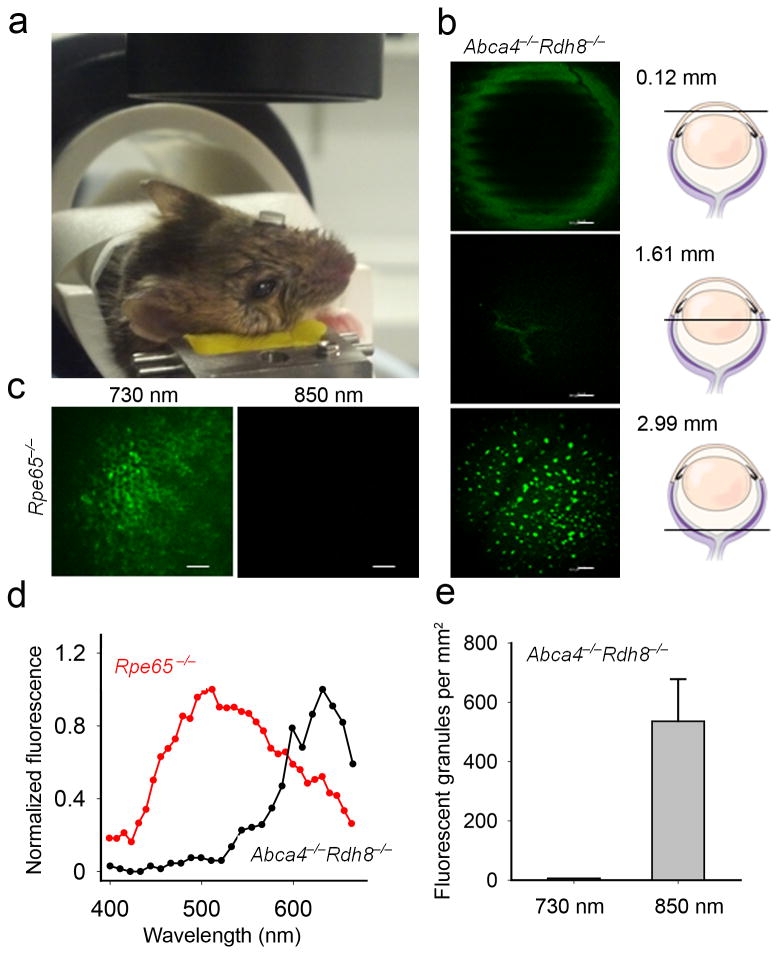
Two-photon excitation microscopy can image retinal molecular processes in vivo. Intrinsically fluorescent retinyl esters in subcellular structures called retinosomes are an integral part of the visual chromophore regeneration pathway. Fluorescent condensation products of all-trans-retinal accumulate in the eye with age and are also associated with age-related macular degeneration (AMD). Here, we report repetitive, dynamic imaging of these compounds in live mice through the pupil of the eye. By leveraging advanced adaptive optics, we developed a data acquisition algorithm that permitted the identification of retinosomes and condensation products in the retinal pigment epithelium by their characteristic localization, spectral properties and absence in genetically modified or drug-treated mice. This imaging approach has the potential to detect early molecular changes in retinoid metabolism that trigger light- and AMD-induced retinal defects and to assess the effectiveness of treatments for these conditions.


Conflict of interest statement

Competing Financial Interests: G.P. and Z.D. are employees of Polgenix. K.P. is CSO at Polgenix Inc. K.P. is an inventor of the U.S. Patent No. 7,706,863 and U.S. Patent No. 8,346,345 whose value may be affected by this publication. N.S.A and M.G., report no conflict of interest. The J.J.H and D.R.W. laboratories received support from Polgenix Inc.

Figures

Figure 1
Figure 1
Two-photon microscopy (TPM) for imaging of mouse retina and RPE. (a) TPM system layout. DC stands for group velocity dispersion pre–compensation; EOM - electro–optic modulator; DM6000 - upright microscope; PMT - photomultiplier tube. (b) Dichroic mirror (DCh) and barrier filter 680 SPET separate fluorescence and excitation light. (c) Layout of the adaptive optics system. FMK1 and FMK2 stand for fold mirrors on kinematic magnetic bases; L1, L2, L3 and L4 - lenses; DM - deformable mirror; FM1, FM2 and FM3 - fold mirrors. (d) Left panel, RPE image in an ex vivo 1-month-old Abca4−/−Rdh8−/− mouse after exposure to bright light, obtained with (top image) and without (bottom image) DC; right panel, mean fluorescence measured with and without DC; error bars indicate S.D, n=3. (e) Upper row, images of the RPE in an ex vivo 3-month-old Rpe65−/− mouse obtained during DM optimization: left, at the start of optimization, with DM in the neutral position; right, at the completion of the imaging session; trial represents an image obtained with non-optimal DM settings; optimal, - an image obtained with DM settings that improved image quality. Bottom row pictures the corresponding DM surfaces. (f) Quantification of image quality, m stands for mean. Scale bars represent 100 μm.
Figure 2
Figure 2
Two–photon images of ex vivo mouse RPE and retina obtained through the mouse eye pupil. Excitation wavelengths and genetic background are listed in each image. (a) The RPE in 3-month-old Rpe65−/− mouse eye. The inset in the right bottom quarter provides a magnified view of the RPE from the area outlined with a white rectangle. (b) The RPE in 6-month-old Abca4−/−Rdh8−/− mouse eye. (c) The RPE in 2-month-old WT mouse eye. (d) The ganglion cell layer in 2-month-old WT mouse eye. White arrows in b and d point to the nuclei. Scale bars represent 50 μm in all panels.
Figure 3
Figure 3
Use of two-photon imaging for ophthalmic drug screening. (a) Ret–NH2 protects RPE of 1-month-old Abca4−/−Rdh8−/− mouse from bright light induced accumulation of fluorescent granules. Representative ex vivo images obtained 7 and 14 days after bright light exposure; images obtained with a ‘through the sclera’ configuration are included for comparison. Excitation with 730 nm was used for the upper row images whereas 850 nm was employed for the lower row. (b) Individual rod photoreceptors expressing rhodopsin-GFP fusion protein are visible in photoreceptor layer of 2-month-old hrhoG/hrhoG mice. (c) Two-photon excited emission spectra from fluorescent granules in the RPE of Abca4−/−Rdh8−/− mouse obtained through the sclera (black) and pupil (red). Spectrum from photoreceptors in hrhoG/hrhoG mice is shown in gray. (d) Quantification of Ret–NH2 impact on accumulation of fluorescent granules in the RPE, based on images as shown in (a); ND stands for none detected; error bars indicate S.D., n = 3. (e) Lower zoom image of the RPE in 6-week-old mouse not treated with Ret–NH2, showing the optic disc is displayed in upper panel. Lower panel shows a magnified view from RPE area outlined with white rectangle in the upper image. Scale bars represent 30 μm in (a, b) and lower panel of (e) and 220 μm in the upper panel of (e).
Figure 4
Figure 4
Set-up for two-photon RPE imaging in living mice. (a) During imaging a contact lens covers mouse eye facing the objective. (b) Representative images of a pigmented 7-week-old Abca4−/−Rdh8−/− mouse eye obtained in vivo with 850 nm excitation 14 days after exposure to bright light, at different depths along Z-axis; a 120 μm section through the cornea, a 1608 μm section showing lens sutures, and a 2987 μm section revealing fluorescent granules in the RPE. (c) Images of the RPE in live albino 7-week-old Rpe65−/− mice obtained with 730 nm and 850 nm excitation. (d) Fluorescence emission spectra from RPE of 7-week-old Abca4−/−Rdh8−/− mice obtained with 850 nm and 7-week-old Rpe65−/− mice obtained with 730 nm excitation light in vivo. (e) Quantification of fluorescent granules. Error bars indicate S.D., n = 3.

Similar articles

Cited by

References

    1. Helmchen F, Denk W. Deep tissue two-photon microscopy. Nat Methods. 2005;2:932–940. - PubMed
    1. Boettner EA, Wolter JR. Transmission of the oculer media. Invest Ophthalmol Vis Sci. 1962;1:776–183.
    1. Imanishi Y, Batten ML, Piston DW, Baehr W, Palczewski K. Noninvasive two-photon imaging reveals retinyl ester storage structures in the eye. J Cell Biol. 2004;164:373–383. - PMC - PubMed
    1. Imanishi Y, Gerke V, Palczewski K. Retinosomes: new insights into intracellular managing of hydrophobic substances in lipid bodies. J Cell Biol. 2004;166:447–453. - PMC - PubMed
    1. Palczewska G, et al. Noninvasive multiphoton fluorescence microscopy resolves retinol and retinal condensation products in mouse eyes. Nat Med. 2010;16:1444–1449. - PMC - PubMed

Publication types