

The antihypertension drug doxazosin inhibits tumor growth and angiogenesis by decreasing VEGFR-2/Akt/mTOR signaling and VEGF and HIF-1α expression
- PMID: 24952732
- PMCID: PMC4148112
- DOI: 10.18632/oncotarget.2064
The antihypertension drug doxazosin inhibits tumor growth and angiogenesis by decreasing VEGFR-2/Akt/mTOR signaling and VEGF and HIF-1α expression
Abstract
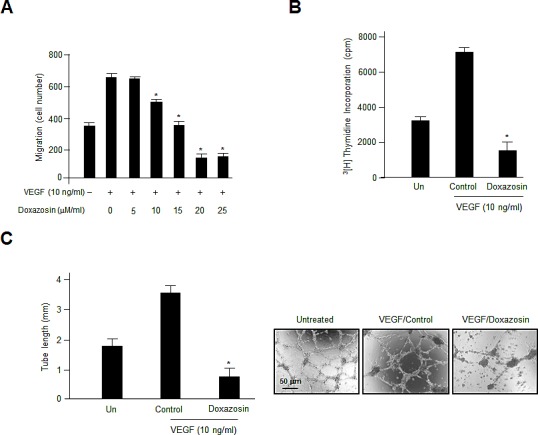
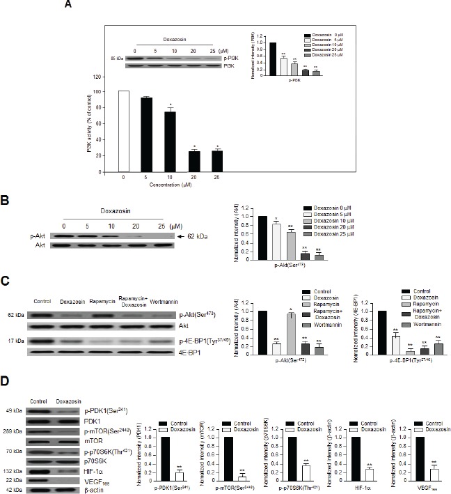
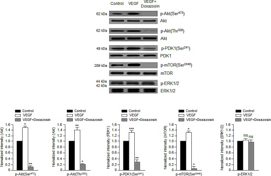
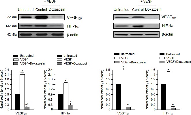
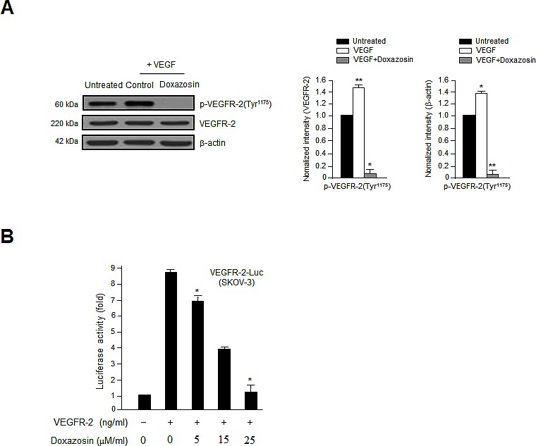
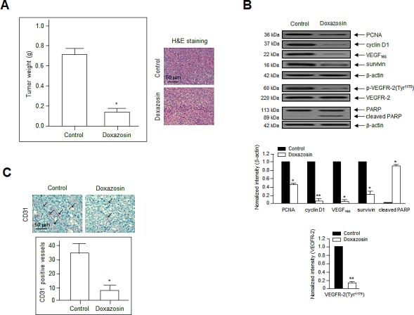
Doxazosin is an α1 adrenergic receptor blocker that also exerts antitumor effects. However, the underlying mechanisms by which it modulates PI3K/Akt intracellular signaling are poorly understood. In this study, we reveal that doxazosin functions as a novel antiangiogenic agent by inhibiting vascular endothelial growth factor (VEGF)-induced cell migration and proliferation. It also inhibited VEGF-induced capillary-like structure tube formation in vitro. Doxazosin inhibited the phosphorylation of VEGF receptor-2 (VEGFR-2) and downstream signaling, including PI3K, Akt, 3-phosphoinositide-dependent protein kinase 1 (PDK1), mammalian target of rapamycin (mTOR), and hypoxia-inducible factor 1 (HIF-1α). However, it had no effect on VEGF-induced extracellular signal-regulated kinase 1/2 (ERK1/2) phosphorylation. Furthermore, doxazosin reduced tumor growth and suppressed tumor vascularization in a xenograft human ovarian cancer model. These results provide evidence that doxazosin functions in the endothelial cell system to modulate angiogenesis by inhibiting Akt and mTOR phosphorylation and interacting with VEGFR-2.
Conflict of interest statement
The authors declare no conflict of interest.
Figures
Similar articles
-
sMEK1 inhibits endothelial cell proliferation by attenuating VEGFR-2-dependent-Akt/eNOS/HIF-1α signaling pathways.Oncotarget. 2015 Oct 13;6(31):31830-43. doi: 10.18632/oncotarget.5570. Oncotarget. 2015. PMID: 26378810 Free PMC article.
-
GABARBP down-regulates HIF-1α expression through the VEGFR-2 and PI3K/mTOR/4E-BP1 pathways.Cell Signal. 2014 Jul;26(7):1506-13. doi: 10.1016/j.cellsig.2014.03.017. Epub 2014 Mar 29. Cell Signal. 2014. PMID: 24686084
-
Magnolol suppresses hypoxia-induced angiogenesis via inhibition of HIF-1α/VEGF signaling pathway in human bladder cancer cells.Biochem Pharmacol. 2013 May 1;85(9):1278-87. doi: 10.1016/j.bcp.2013.02.009. Epub 2013 Feb 14. Biochem Pharmacol. 2013. PMID: 23416116
-
Dual Roles of the AMP-Activated Protein Kinase Pathway in Angiogenesis.Cells. 2019 Jul 19;8(7):752. doi: 10.3390/cells8070752. Cells. 2019. PMID: 31331111 Free PMC article. Review.
-
Emerging roles of post-translational modifications in signal transduction and angiogenesis.Proteomics. 2015 Jan;15(2-3):300-9. doi: 10.1002/pmic.201400183. Epub 2014 Oct 8. Proteomics. 2015. PMID: 25161153 Free PMC article. Review.
Cited by
-
The role of hypoxia-inducible factor-1α in radiation-induced autophagic cell death in breast cancer cells.Tumour Biol. 2015 Sep;36(9):7077-83. doi: 10.1007/s13277-015-3425-z. Epub 2015 Apr 15. Tumour Biol. 2015. PMID: 25874499
-
Doxazosin inhibits vasculogenic mimicry in human non‑small cell lung cancer through inhibition of the VEGF‑A/VE‑cadherin/mTOR/MMP pathway.Oncol Lett. 2024 Feb 22;27(4):170. doi: 10.3892/ol.2024.14303. eCollection 2024 Apr. Oncol Lett. 2024. PMID: 38455663 Free PMC article.
-
ZLM-7 exhibits anti-angiogenic effects via impaired endothelial cell function and blockade of VEGF/VEGFR-2 signaling.Oncotarget. 2016 Apr 5;7(14):19018-30. doi: 10.18632/oncotarget.7968. Oncotarget. 2016. PMID: 26967559 Free PMC article.
-
EMMPRIN/CD147 is a novel coreceptor of VEGFR-2 mediating its activation by VEGF.Oncotarget. 2015;6(12):9766-80. doi: 10.18632/oncotarget.2870. Oncotarget. 2015. PMID: 25825981 Free PMC article.
-
Insights into cardio-oncology: Polypharmacology of quinazoline-based α1-adrenoceptor antagonists.World J Cardiol. 2015 May 26;7(5):238-42. doi: 10.4330/wjc.v7.i5.238. World J Cardiol. 2015. PMID: 26015856 Free PMC article. Review.
References
-
- Folkman J, Shing Y. Angiogenesis. J. Biol. Chem. 1992;267:10931–10934. - PubMed
-
- Risau W. Mechanisms of angiogenesis. Nature. 1997;386:671–674. - PubMed
-
- Ferrara N. VEGF and the quest for tumour angiogenesis factors. Nat. Rev. Cancer. 2002;2:795–803. - PubMed
-
- Yoshiji H, Gomez DE, Shibuya M, Thorgeirsson UP. Expression of vascular endothelial growth factor, its receptor, and other angiogenic factors in human breast cancer. Cancer Res. 1996;56:2013–6. - PubMed
-
- Gavin TP, Robinson CB, Yeager RC, England JA, Nifong LW, Hickner RC. Angiogenic growth factor response to acute systemic exercise in human skeletal muscle. J. Appl. Physiol. 2004;96:19–24. - PubMed
Publication types
MeSH terms
Substances
LinkOut - more resources
Full Text Sources
Other Literature Sources
Medical
Molecular Biology Databases
Miscellaneous
