

Ordering of mutations in preinvasive disease stages of esophageal carcinogenesis
- PMID: 24952744
- PMCID: PMC4116294
- DOI: 10.1038/ng.3013
Ordering of mutations in preinvasive disease stages of esophageal carcinogenesis
Abstract
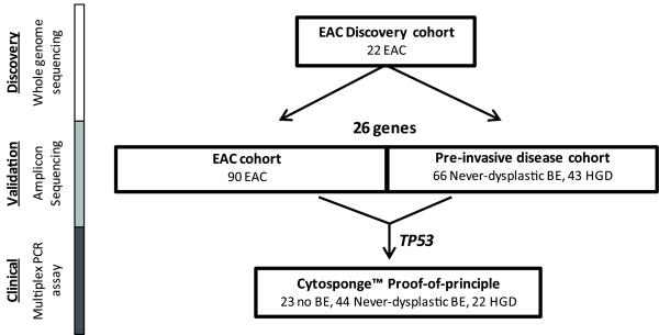
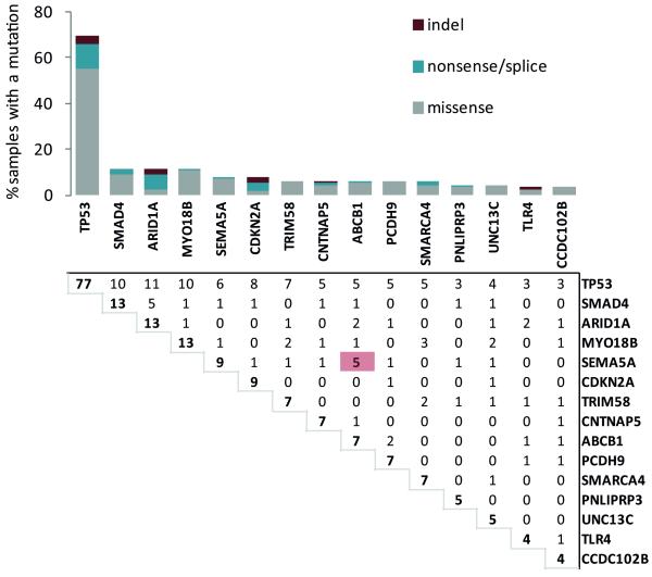
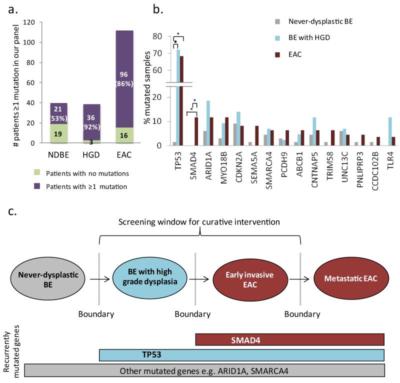
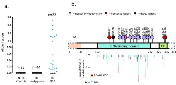
Cancer genome sequencing studies have identified numerous driver genes, but the relative timing of mutations in carcinogenesis remains unclear. The gradual progression from premalignant Barrett's esophagus to esophageal adenocarcinoma (EAC) provides an ideal model to study the ordering of somatic mutations. We identified recurrently mutated genes and assessed clonal structure using whole-genome sequencing and amplicon resequencing of 112 EACs. We next screened a cohort of 109 biopsies from 2 key transition points in the development of malignancy: benign metaplastic never-dysplastic Barrett's esophagus (NDBE; n=66) and high-grade dysplasia (HGD; n=43). Unexpectedly, the majority of recurrently mutated genes in EAC were also mutated in NDBE. Only TP53 and SMAD4 mutations occurred in a stage-specific manner, confined to HGD and EAC, respectively. Finally, we applied this knowledge to identify high-risk Barrett's esophagus in a new non-endoscopic test. In conclusion, mutations in EAC driver genes generally occur exceptionally early in disease development with profound implications for diagnostic and therapeutic strategies.
Figures
Comment in
-
Genetics. Driver genes are mutated early in the course of oesophageal adenocarcinoma.Nat Rev Gastroenterol Hepatol. 2014 Aug;11(8):453. doi: 10.1038/nrgastro.2014.117. Epub 2014 Jul 15. Nat Rev Gastroenterol Hepatol. 2014. PMID: 25023036 No abstract available.
-
Genetics: Driver genes are mutated early in the course of oesophageal adenocarcinoma.Nat Rev Clin Oncol. 2014 Aug;11(8):439. doi: 10.1038/nrclinonc.2014.121. Epub 2014 Jul 15. Nat Rev Clin Oncol. 2014. PMID: 25027592 No abstract available.
-
From genomics to diagnostics of esophageal adenocarcinoma.Nat Genet. 2014 Aug;46(8):806-7. doi: 10.1038/ng.3047. Nat Genet. 2014. PMID: 25070798 Free PMC article. No abstract available.
References
Publication types
MeSH terms
Supplementary concepts
Grants and funding
- 12088/CRUK_/Cancer Research UK/United Kingdom
- 11906/CRUK_/Cancer Research UK/United Kingdom
- 05/12/01/DH_/Department of Health/United Kingdom
- MC_U105365007/MRC_/Medical Research Council/United Kingdom
- A12770/CRUK_/Cancer Research UK/United Kingdom
- NIHR-RP-02-12-011/DH_/Department of Health/United Kingdom
- WT_/Wellcome Trust/United Kingdom
- 20240/CRUK_/Cancer Research UK/United Kingdom
- 16942/CRUK_/Cancer Research UK/United Kingdom
- MC_UU_12022/2/MRC_/Medical Research Council/United Kingdom
- G1002565/MRC_/Medical Research Council/United Kingdom
- 15874/CRUK_/Cancer Research UK/United Kingdom
- 14545/CRUK_/Cancer Research UK/United Kingdom
LinkOut - more resources
Full Text Sources
Other Literature Sources
Medical
Molecular Biology Databases
Research Materials
Miscellaneous
