

RFX1-dependent activation of SHP-1 induces autophagy by a novel obatoclax derivative in hepatocellular carcinoma cells
- PMID: 24952874
- PMCID: PMC4148109
- DOI: 10.18632/oncotarget.2054
RFX1-dependent activation of SHP-1 induces autophagy by a novel obatoclax derivative in hepatocellular carcinoma cells
Abstract
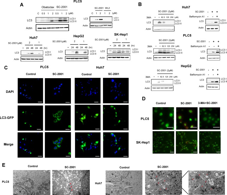
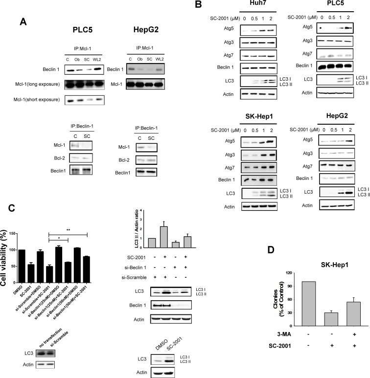
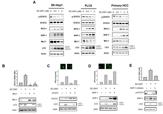
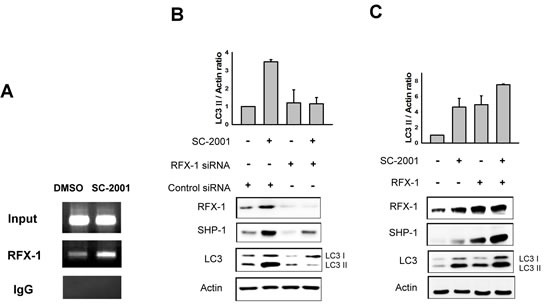
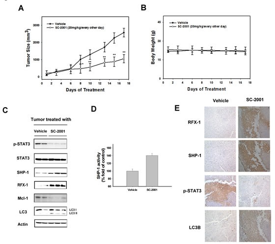
Obatoclax is a small molecule which targets the Bcl-2 family, and is to treat leukemia, lymphoma and lung carcinoma. Previously, an obatoclax analogue, SC-2001, was found to disrupt the protein-protein interactions of the Bcl-2 family and also repress Bcl-XL and Mcl-1 expression via STAT3 inactivation. Here, we report a novel mechanism of autophagy induction by SC-2001 in liver cancer cells. The findings indicate that SC-2001 induced the autophagy marker LC3-II in four hepatocellular carcinoma (HCC) cells. Autophagosomes induced by SC-2001-treated cells were confirmed by electron microscopy. SC-2001 activated SHP-1, dephosphorylated STAT3 and Mcl-1, and subsequently released free beclin 1. Overexpression of STAT3 and Mcl-1 in PLC5 cells attenuated the induction of SC-2001 on autophagy. Abolishment of SHP-1 by a specific inhibitor aboragated the autophagic effects induced by SC-2001. In addition, it was further revealed that RFX-1, a transcription factor of SHP-1, is a critical regulator in SC-2001-mediated autophagy. Downregulation of RFX-1 by si-RNA protected cells from SC-2001-induced autophagy. Importantly, Huh7 tumor-bearing nude mice treated with SC-2001 showed downregulation of Mcl-1 and p-STAT3 protein expression and upregulation of SHP-1, LC3II, and RFX-1 protein expression. In summary, our data suggest that SC-2001 induces autophagic cell death in a RFX1/SHP-1/STAT3/Mcl-1 signaling cascade.
Conflict of interest statement
The authors have nothing relevant to this manuscript to disclose.
Figures
Similar articles
-
Mcl-1-dependent activation of Beclin 1 mediates autophagic cell death induced by sorafenib and SC-59 in hepatocellular carcinoma cells.Cell Death Dis. 2013 Feb 7;4(2):e485. doi: 10.1038/cddis.2013.18. Cell Death Dis. 2013. PMID: 23392173 Free PMC article.
-
Obatoclax analog SC-2001 inhibits STAT3 phosphorylation through enhancing SHP-1 expression and induces apoptosis in human breast cancer cells.Breast Cancer Res Treat. 2014 Jul;146(1):71-84. doi: 10.1007/s10549-014-3000-0. Epub 2014 Jun 6. Breast Cancer Res Treat. 2014. PMID: 24903225
-
SC-2001 overcomes STAT3-mediated sorafenib resistance through RFX-1/SHP-1 activation in hepatocellular carcinoma.Neoplasia. 2014 Jul;16(7):595-605. doi: 10.1016/j.neo.2014.06.005. Epub 2014 Jul 18. Neoplasia. 2014. PMID: 25047655 Free PMC article.
-
A novel obatoclax derivative, SC-2001, induces apoptosis in hepatocellular carcinoma cells through SHP-1-dependent STAT3 inactivation.Cancer Lett. 2012 Aug 1;321(1):27-35. doi: 10.1016/j.canlet.2012.03.023. Epub 2012 Mar 28. Cancer Lett. 2012. PMID: 22465052
-
RFX-1-dependent activation of SHP-1 inhibits STAT3 signaling in hepatocellular carcinoma cells.Carcinogenesis. 2014 Dec;35(12):2807-14. doi: 10.1093/carcin/bgu210. Epub 2014 Oct 16. Carcinogenesis. 2014. PMID: 25322871
Cited by
-
Sorafenib analogue SC-60 induces apoptosis through the SHP-1/STAT3 pathway and enhances docetaxel cytotoxicity in triple-negative breast cancer cells.Mol Oncol. 2017 Mar;11(3):266-279. doi: 10.1002/1878-0261.12033. Epub 2017 Feb 7. Mol Oncol. 2017. PMID: 28084011 Free PMC article.
-
Protective Effect of Curcumin on Bone Trauma in a Rat Model via Expansion of Myeloid Derived Suppressor Cells.Med Sci Monit. 2020 Nov 13;26:e924724. doi: 10.12659/MSM.924724. Med Sci Monit. 2020. PMID: 33184252 Free PMC article.
-
Roles of protein tyrosine phosphatases in hepatocellular carcinoma progression (Review).Oncol Rep. 2023 Mar;49(3):48. doi: 10.3892/or.2023.8485. Epub 2023 Jan 20. Oncol Rep. 2023. PMID: 36660927 Free PMC article. Review.
-
RFX1: a promising therapeutic arsenal against cancer.Cancer Cell Int. 2021 May 8;21(1):253. doi: 10.1186/s12935-021-01952-6. Cancer Cell Int. 2021. PMID: 33964962 Free PMC article. Review.
-
Systematic analyses and comprehensive field synopsis of genetic association studies in hepatocellular carcinoma.Oncotarget. 2016 Jul 19;7(29):45757-45763. doi: 10.18632/oncotarget.9937. Oncotarget. 2016. PMID: 27304192 Free PMC article. Review.
References
-
- Levine B, Klionsky DJ. Development by self-digestion: molecular mechanisms and biological functions of autophagy. Dev Cell. 2004;6(4):463–477. - PubMed
-
- Mizushima N. Autophagy: process and function. Genes Dev. 2007;21(22):2861–2873. - PubMed
-
- Maiuri MC, Zalckvar E, Kimchi A, Kroemer G. Self-eating and self-killing: crosstalk between autophagy and apoptosis. Nat Rev Mol Cell Biol. 2007;8(9):741–752. - PubMed
-
- Codogno P, Mehrpour M, Proikas-Cezanne T. Canonical and non-canonical autophagy: variations on a common theme of self-eating? Nat Rev Mol Cell Biol. 2012;13(1):7–12. - PubMed
Publication types
MeSH terms
Substances
LinkOut - more resources
Full Text Sources
Other Literature Sources
Research Materials
Miscellaneous
