. 2014 Jun 22:14:463.
doi: 10.1186/1471-2407-14-463.

FHL1C induces apoptosis in Notch1-dependent T-ALL cells through an interaction with RBP-J

Affiliations

FHL1C induces apoptosis in Notch1-dependent T-ALL cells through an interaction with RBP-J

Wei Fu et al. BMC Cancer. .

Abstract

Background: Aberrantly activated Notch signaling has been found in more than 50% of patients with T-cell acute lymphoblastic leukemia (T-ALL). Current strategies that employ γ-secretase inhibitors (GSIs) to target Notch activation have not been successful. Many limitations, such as non-Notch specificity, dose-limiting gastrointestinal toxicity and GSI resistance, have prompted an urgent need for more effective Notch signaling inhibitors for T-ALL treatment. Human four-and-a-half LIM domain protein 1C (FHL1C) (KyoT2 in mice) has been demonstrated to suppress Notch activation in vitro, suggesting that FHL1C may be new candidate target in T-ALL therapy. However, the role of FHL1C in T-ALL cells remained unclear.

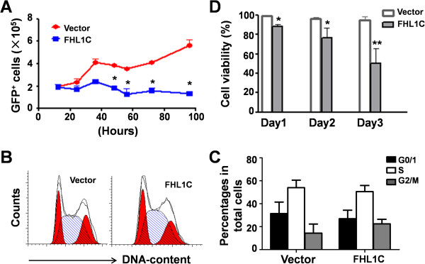
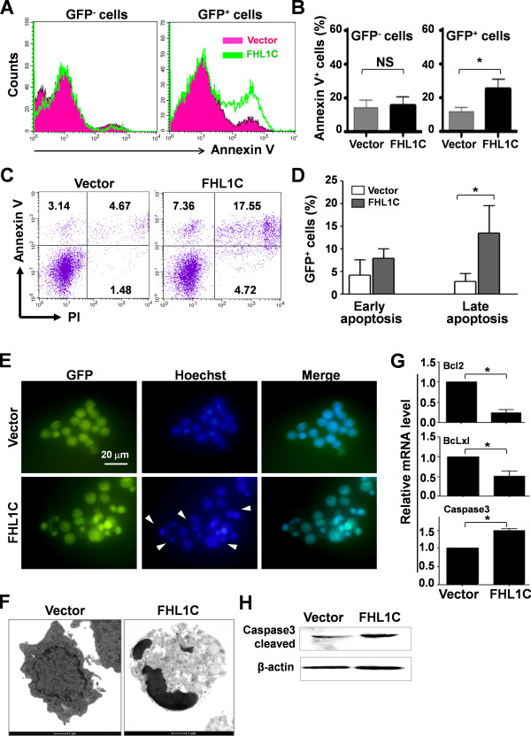
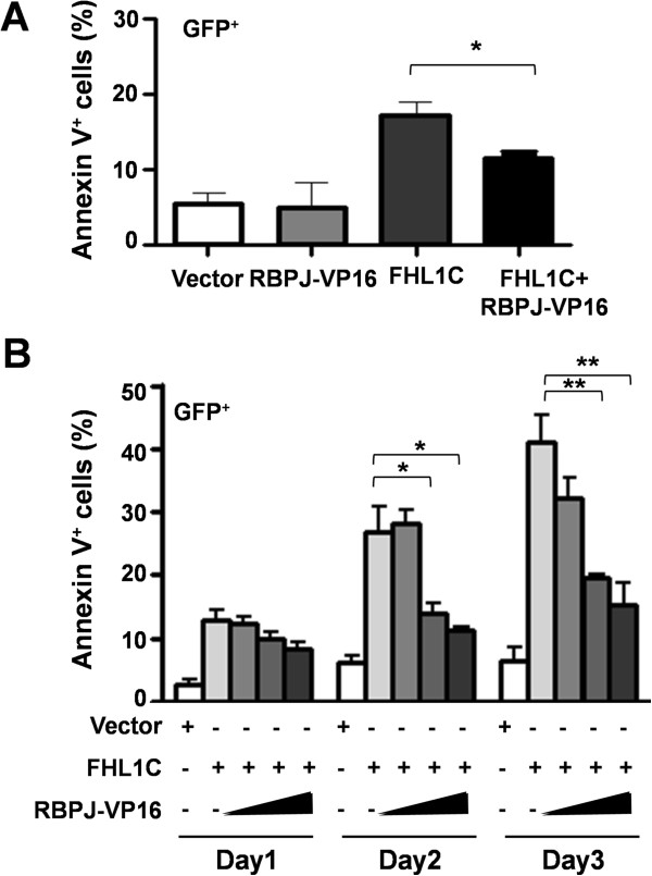
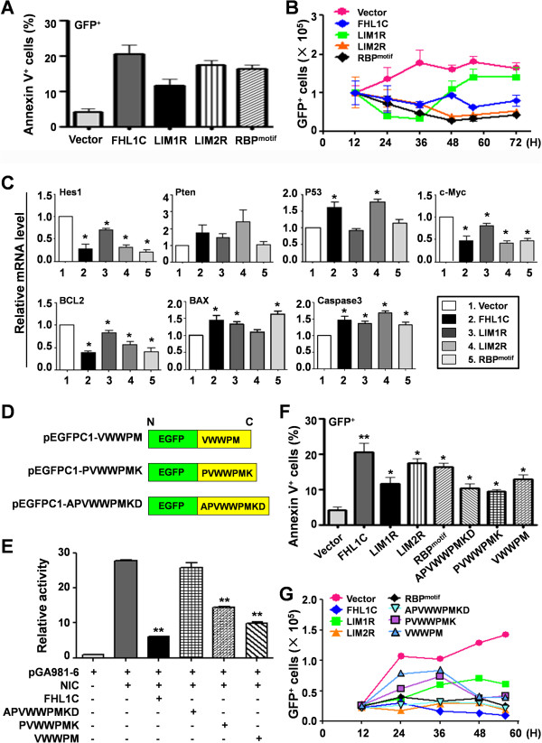
Methods: Using RT-PCR, we amplified full-length human FHL1C, and constructed full-length and various truncated forms of FHL1C. Using cell transfection, flow cytometry, transmission electron microscope, real-time RT-PCR, and Western blotting, we found that overexpression of FHL1C induced apoptosis of Jurkat cells. By using a reporter assay and Annexin-V staining, the minimal functional sequence of FHL1C inhibiting RBP-J-mediated Notch transactivation and inducing cell apoptosis was identified. Using real-time PCR and Western blotting, we explored the possible molecular mechanism of FHL1C-induced apoptosis. All data were statistically analyzed with the SPSS version 12.0 software.

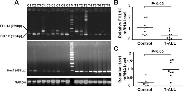
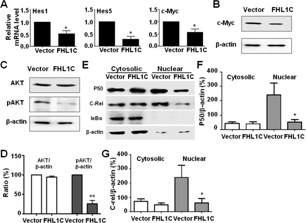
Results: In Jurkat cells derived from a Notch1-associated T-ALL cell line insensitive to GSI treatment, we observed that overexpression of FHL1C, which is down-regulated in T-ALL patients, strongly induced apoptosis. Furthermore, we verified that FHL1C-induced apoptosis depended on the RBP-J-binding motif at the C-terminus of FHL1C. Using various truncated forms of FHL1C, we found that the RBP-J-binding motif of FHL1C had almost the same effect as full-length FHL1C on the induction of apoptosis, suggesting that the minimal functional sequence in the RBP-J-binding motif of FHL1C might be a new drug candidate for T-ALL treatment. We also explored the molecular mechanism of FHL1C overexpression-induced apoptosis, which suppressed downstream target genes such as Hes1 and c-Myc and key signaling pathways such as PI3K/AKT and NF-κB of Notch signaling involved in T-ALL progression.

Conclusions: Our study has revealed that FHL1C overexpression induces Jurkat cell apoptosis. This finding may provide new insights in designing new Notch inhibitors based on FHL1C to treat T-ALL.


Figures

Figure 1
Figure 1
Expression of FHL1C and Hes1 was detected in T-ALL patients and healthy donors. (A) RT-PCR analysis of FHL1C and Hes1 mRNA in PBMCs from 8 T-ALL patients and 9 healthy donors, with GAPDH as an internal control. (B, C) Relative mRNA levels of FHL1C (B) and Hes1 (C) to GAPDH in PBMCs from T-ALL patients and controls were compared. The horizontal lines indicated median expression levels.
Figure 2
Figure 2
Overexpression of FHL1C in Jurkat cells resulted in reduced cell viability. (A) Jurkat cells (5 × 106) were transfected with pEGFP or pEGFP-FHL1C by using the Nucleofection method. The numbers of viable EGFP-positive cells were determined every 12 h by cell counting and FACS analysis. (B, C) Cell cycle progression of Jurkat cells in (A) was determined 48 h post-transfection by FACS after PI staining (B). Cells in each phase were compared between the two groups (C). (D) Total viability of Jurkat cells transfected in (A) was monitored by using trypan blue exclusion assay. Bars = means ± S.D (n = 3), *P < 0.05, **P < 0.01.
Figure 3
Figure 3
FHL1C overexpression induced apoptosis in Jurkat cells. (A) Jurkat cells were transiently transfected with pEGFP or pEGFP-FHL1C by using the Nucleofection method. Apoptosis in the GFP and GFP+ fractions of cells was determined by AnnexinV staining followed by FACS 48 h post-transfection. (B) Percentages of apoptotic cells (Annexin V+) in GFP and GFP+ cell fractions in (A) were compared. (C) Jurkat cells were transfected with pEGFP or pEGFP-FHL1C by using the Nucleofection method. Early and late apoptotic cells were depicted 48 h post-transfection by using Annexin V and PI staining followed by FACS. (D) GFP+ cells in early and late apoptotic phases in (C) were compared. (E) Jurkat cells were transiently transfected with pEGFP or pEGFP-FHL1C by using the Nucleofection method. Cells were stained with Hoechst 24 h post-transfection and nuclei were observed under a fluorescence microscope. Arrow heads indicate Hoechst-positive apoptotic nuclei. (F) Typical cell apoptosis in (E) was depicted under TEM. Intact cell membrane, organelles and normal nuclear morphology were observed in vector-transfected cells, whereas incomplete membrane and condensed nuclei were observed in cells overexpressing FHL1C (magnification, × 9900). (G) Total RNA was prepared from cells in (E) 24 h post-transfection. The mRNA levels of the apoptosis-related molecules were determined by real time RT-PCR, with β-actin as a reference. (H) Cell lysates were prepared from cells in (E) 24 h post-transfection. The level of Caspase3 was determined by Western blot analysis. Bars = means ± S.D (n = 3), *P < 0.05; NS, not significant.
Figure 4
Figure 4
FHL1C induced apoptosis of Jurkat cells through repressing RBP-J. (A) Jurkat cells were transiently transfected with pEGFP, pCMX-VP16-RBP-J, pEGFP-FHL1C or pEGFP-FHL1C plus pCMX-VP16-RBP-J. The percentage of apoptotic (Annexin V+) cells in EGFP+ cell population was measured 24 h after transfection. (B) Constitutively active RBP-J blocked FHL1C-induced apoptosis in Jurkat cells. Jurkat cells were transiently transfected with 1 μg of pEGFP-FHL1C alone or in combination with increasing amounts (0.2, 0.5, 1.0 μg) of pCMX-VP16-RBP-J. Cell apoptosis was measured by Annexin V staining on different days after transfection. The percentages of apoptotic (Annexin V+) cells in EGFP+ cell population were shown. Bars = means ± S.D (n = 3), *P < 0.05, **P < 0.01.
Figure 5
Figure 5
The RBP-J-binding motif was sufficient to induce apoptosis in Jurkat cells. (A) Full length and differentially truncated FHL1C (Additional file 7: Figure S3A) were inserted into pEGFPC1 in frame, and were used to transfect Jurkat cells. The cells were analyzed by Annexin V staining followed by FACS 48 h post-transfection. The percentages of apoptotic (Annexin V+) cells in the EGFP+ cell population were determined. (B) Jurkat cells were transiently transfected with plasmids as in (A). The numbers of EGFP+ cells were counted at different time points after transfection. (C) Jurkat cells were transiently transfected with plasmids as in (A). Cells were harvested 48 h post-transfection for RNA extraction. The mRNA expression levels of Hes1, Pten, Myc, p53, Bcl2, Bax, and Caspase3 were detected by qRT-PCR, with β-actin as a reference. (D) The core sequences with different length of the RBP-J-binding motif in FHL1C were fused to the 3′ terminus of EGFP in frame, to construct plasmids expressing EGFP with RBP-J-binding motif at the C-terminus. (E) EGFP containing RBP-J-binding motif inhibited NIC-mediated transactivation of RBP-J specific reporter construct. HeLa cells were transfected with different plasmids as indicated, and luciferase activity in the cell lysates was examined 48 h after transfection. (F) Jurkat cells were transiently transfected with plasmids as indicated. The cells were analyzed by Annexin V staining followed by FACS 48 h after the transfection. The percentages of apoptotic (Annexin V+) cells in the EGFP+ cell population were determined. (G) Jurkat cells were transiently transfected with plasmids as indicated. The numbers of GFP+ cells were counted at different time points after transfection. Bars = means ± S.D (n = 3), *P < 0.05, **P < 0.01.
Figure 6
Figure 6
Overexpression of FHL1C induced apoptosis of Jurkat cells involving multiple effectors and pathways. (A) Jurkat cells were transfected with pEGFP or pEGFP-FHL1C by using the Nucleofection method. The cells were harvested 48 h post-transfection, and the mRNA levels of Hes1, Hes5 and c-Myc were detected by real time RT-PCR, with β-actin as a reference. (B) Jurkat cells were transfected as in (A). The protein level of c-Myc was determined by using Western blotting. (C,D) Cell lysates were prepared from Jurkat cells transfected with pEGFP or pEGFP-FHL1C for 48 h. AKT and phosphorylated AKT (pAKT) were analyzed by Western blotting (C). The relative levels of AKT and pAKT were quantified and compared, with β-actin as an internal control (D). (E-G) Jurkat cells were transfected with pEGFP or pEGFP-FHL1C by using the Nucleofection method. Cells were harvested 24 h post-transfection, and the cytosolic and nuclear extracts were fractioned. P50, c-Rel and IκB were determined by Western blotting (E). The relative levels of P50 (F) and c-Rel (G) were quantified and compared, with β-actin as an internal control. Bars = means ± S.D (n = 3), *P < 0.05, **P < 0.01.

Similar articles

Cited by

References

    1. Osanyingbemi-Obidi J, Dobromilskaya I, Illei PB, Hann CL, Rudin CM. Notch signaling contributes to lung cancer clonogenic capacity in vitro but may be circumvented in tumorigenesis in vivo. Molecular cancer research: MCR. 2011;9(12):1746–1754. - PMC - PubMed
    1. Shao L, Wang Y, Chang J, Luo Y, Meng A, Zhou D. Hematopoietic stem cell senescence and cancer therapy-induced long-term bone marrow injury. Translational cancer research. 2013;2(5):397–411. - PMC - PubMed
    1. Wang L, Cheng T, Zheng G. The impact of tumor microenvironments on stem cells. Translational cancer research. 2013;2(5):422–428.
    1. Kopan R, Ilagan MX. The canonical notch signaling pathway: unfolding the activation mechanism. Cell. 2009;137(2):216–233. - PMC - PubMed
    1. Gridley T. Kick it up a notch: NOTCH1 activation in T-ALL. Cancer Cell. 2004;6(5):431–432. - PubMed

Publication types

MeSH terms

Substances

LinkOut - more resources