. 2014 Aug 1;289(31):21230-41.
doi: 10.1074/jbc.M114.565010. Epub 2014 Jun 21.

Synergic role of nucleophosmin three-helix bundle and a flanking unstructured tail in the interaction with G-quadruplex DNA

Affiliations

Synergic role of nucleophosmin three-helix bundle and a flanking unstructured tail in the interaction with G-quadruplex DNA

Alessandro Arcovito et al. J Biol Chem. .

Abstract

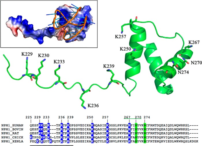
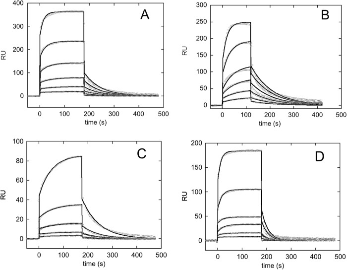
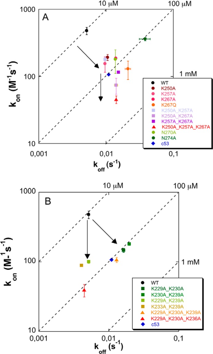
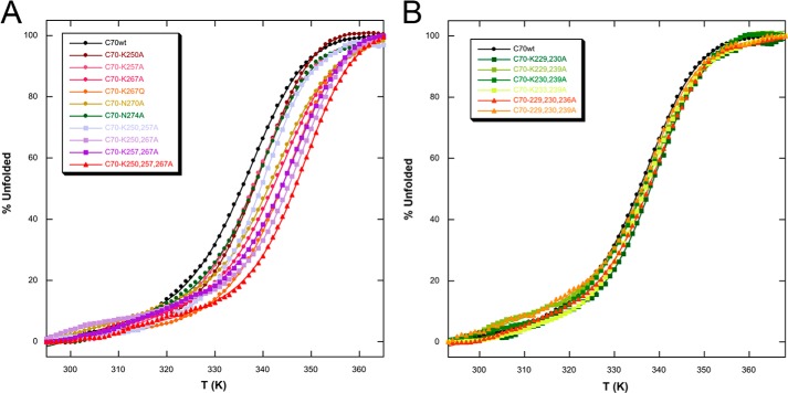
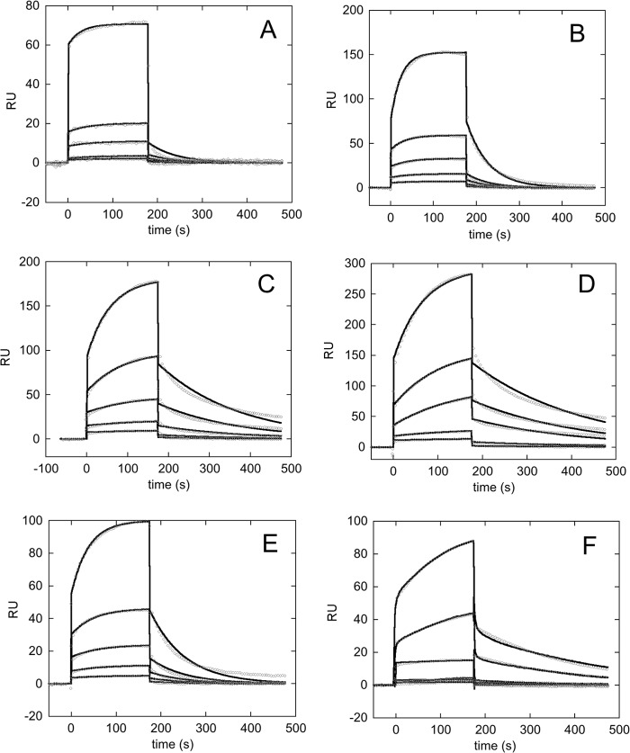
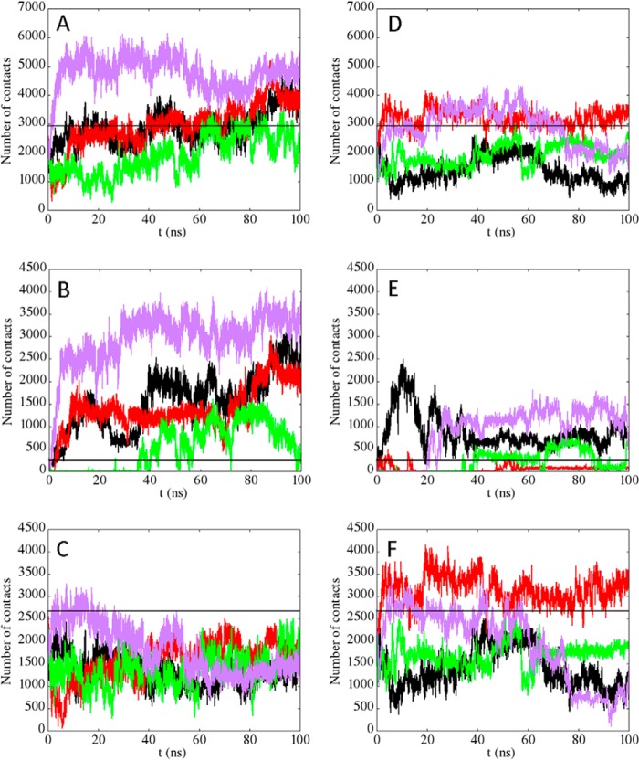
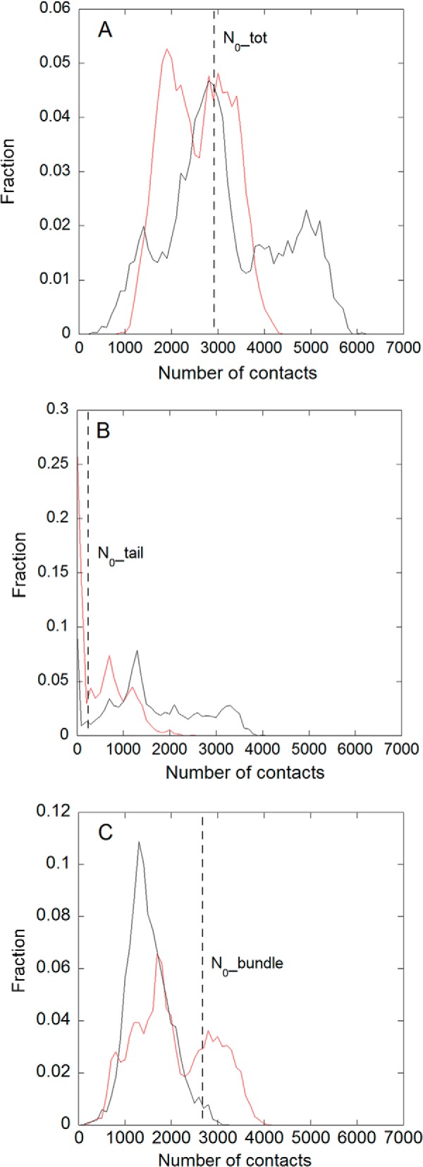
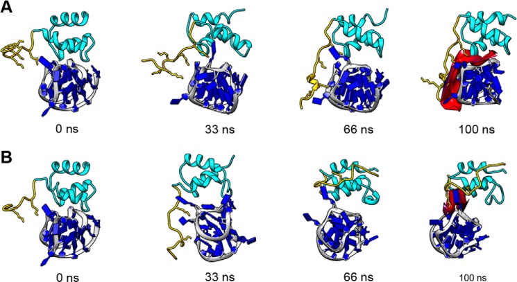
Nucleophosmin (NPM1) is a nucleocytoplasmic shuttling protein, mainly localized at nucleoli, that plays a number of functions in ribosome biogenesis and export, cell cycle control, and response to stress stimuli. NPM1 is the most frequently mutated gene in acute myeloid leukemia; mutations map to the C-terminal domain of the protein and cause its denaturation and aberrant cytoplasmic translocation. NPM1 C-terminal domain binds G-quadruplex regions at ribosomal DNA and at gene promoters, including the well characterized sequence from the nuclease-hypersensitive element III region of the c-MYC promoter. These activities are lost by the leukemic variant. Here we analyze the NPM1/G-quadruplex interaction, focusing on residues belonging to both the NPM1 terminal three-helix bundle and a lysine-rich unstructured tail, which has been shown to be necessary for high affinity recognition. We performed extended site-directed mutagenesis and measured binding rate constants through surface plasmon resonance analysis. These data, supported by molecular dynamics simulations, suggest that the unstructured tail plays a double role in the reaction mechanism. On the one hand, it facilitates the formation of an encounter complex through long range electrostatic interactions; on the other hand, it directly contacts the G-quadruplex scaffold through multiple and transient electrostatic interactions, significantly enlarging the contact surface.

Keywords: Encounter Complex; Flanking Fuzziness; Intrinsically Disordered Protein; Leukemia; Molecular Dynamics; Protein-DNA Interaction; Surface Plasmon Resonance (SPR).


Figures

FIGURE 1.
FIGURE 1.
The figure shows the structure of NPM1-C70WT, highlighting the terminal three-helix bundle preceded by a lysine-rich unstructured tail (underlined residues in the alignment) that is necessary for high affinity recognition of G-quadruplex oligonucleotides. The three-helix bundle residues found to contact the pu24I oligo as well as the five lysine residues in the tail are shown in sticks. The inset shows the structure of the C70WT·pu24I complex as obtained by experimentally constrained molecular docking (19). The alignment shows that three tail lysines are completely conserved. When not conserved, the total charge of the tail is nevertheless maintained by the presence of nearby lysines.
FIGURE 2.
FIGURE 2.
Sensorgrams of the interaction between the immobilized biotinylated pu24I oligo and selected NPM1 C-terminal domain constructs used as analytes. Experimental data at increasing analyte concentrations are reported in circles, whereas fits to a 1:1 binding model are reported as black lines. A, C70WT was used as the analyte, and concentrations measured were 125, 62.5, 31.25, 15.625, 7.8125, and 3.9 μm. B, C53WT was used as the analyte, and concentrations measured were 125, 62.5, 31.25, 15.625, 7.8125, and 3.9 μm. C, the single bundle mutant C70-N274A was used as the analyte, and concentrations measured were 125, 62.5, 31.25, 15.625, and 7.8125 μm. D, the triple bundle mutant C70-K250A,K257A,K267A was used as the analyte, and concentrations measured were 125, 62.5, 31.25, 15.625, 7.8125, and 3.9 μm. RU, resonance units.
FIGURE 3.
FIGURE 3.
Isoaffinity graphs of the effect of C70 mutations on pu24I binding. For each mutant, the association rate constant kon is reported as a function of the corresponding koff value. Dashed lines represent isoaffinity points at the reported KD values. A, three-helix bundle mutants. Single alanine mutations exert their effect both in terms of reduced kon and increased koff (upper arrow), showing that they contribute to both the formation of the encounter complex and stabilization of the final complex. Residue Asn-274 constitutes a remarkable exception because its alanine mutant has a considerably reduced affinity for the G-quadruplex that is almost entirely due to an ∼10-fold higher koff with respect to WT. The additional effect played by double and triple mutants is exerted primarily in terms of further reduced kon (lower arrow), thus impacting complex formation rather than stabilization. B, tail mutants. For some of the mutants, variations are observed not only for the kon values, as expected in a classical fly casting mechanism, but also in terms of increased koff values, suggesting a direct involvement of the tail in complex stabilization. In both panels, error bars (S.E.) are explicitly shown; when error bars are not visible, it means that they are shorter than the size of the marker.
FIGURE 4.
FIGURE 4.
Circular dichroism analysis of C70 mutants. A, thermal denaturation profiles for C70WT and three-helix bundle mutants. Data highlight that thermodynamic stability is not decreased by mutations. Conversely, for double and triple mutants, an increase in stability is observed. The corresponding Tm values are reported in Table 2. B, thermal denaturation profiles for C70WT and tail mutants. Thermodynamic stability is not affected by mutations. The corresponding Tm values are reported in Table 4. In both panels, straight lines represent best fits of experimental data according to Swint and Robertson (20), and the color code is the same as in Fig. 3.
FIGURE 5.
FIGURE 5.
Sensorgrams of the interaction between the immobilized biotinylated pu24I oligo and C70 tail mutants used as the analytes. Experimental data at increasing analyte concentrations are reported in circles, whereas fits to a 1:1 binding model are reported as black lines. Concentrations used were 125, 62.5, 31.25, 15.625, and 7.8125 μm for all mutants tested. A, C70-K229A,K230A mutant was used as the analyte. B, C70-K230A,K239A mutant was used as the analyte. C, C70-K229A,K239A mutant was used as the analyte. D, C70-K233A,K239A mutant was used as the analyte. E, the triple mutant C70-K229A,K230A,K239A was used as the analyte. F, the triple mutant C70-K229A,K230A,K236A was used as the analyte. RU, resonance units.
FIGURE 6.
FIGURE 6.
Analysis of the trajectories obtained for C70WT and the triple mutant C70-K229A,K230A,K236A. Four independent MD simulations are shown in both cases. A–C, the number of contacts (distances below 6 Å) between C70WT and the pu24I G-quadruplex is reported as a function of time (A, total number of contacts; B, number of contacts from the tail, i.e. residues 225–242; C, number of contacts from the bundle, i.e. residues 243–294). D–F, the number of contacts formed by the triple mutant with the pu24I G-quadruplex are shown (D, total number of contacts; E, number of contacts from the tail; F, number of contacts from the bundle). The black straight lines in the panels represent the total number of contacts in the initial model (N0_tot; A and D), the number of contacts from the tail in the initial model (N0_tail; B and E), and the number of contacts from the bundle in the initial model (N0_bundle; C and F), respectively.
FIGURE 7.
FIGURE 7.
Distributions of the number of contacts in the overall set of MD simulations for the wild type (black line) and the C70-K229A,K230A,K236A mutant (red line). A, total number of contacts. B, number of contacts from the tail, i.e. residues 225–242. C, number of contacts from the bundle, i.e. residues 243–294. The dashed lines in the different panels represent the value of the corresponding number of contacts (A, total, B, tail; C, bundle) in the initial model.
FIGURE 8.
FIGURE 8.
Ribbon representation of the complex structures along the molecular dynamics simulations. A, simulation of the C70WT·pu24I complex corresponding to the black curve of Fig. 6, A–C (wild type). As a starting structure, the structure reported in Gallo et al. (19) was used. Snapshots of the simulation at 0, 33, 66, and 100 ns (from left to right) are reported. The pu24I G-quadruplex is shown in blue. The C70WT three-helix bundle is shown in cyan, and its unstructured tail is shown in yellow (lysine side chains are in sticks). The contact surface at 100 ns between C70WT and pu24I is shown in red, highlighting the role of the tail in stabilizing the complex. B, simulations of the C70-K229A,K230A,K236A·pu24I complex corresponding to the black curve of Fig. 6, D–F. The C70-K229A,K230A,K236A mutant was obtained by in silico substitution, and its interaction with pu24I was simulated using the same starting structure as in A. Four snapshots at the same time points as in A are shown. The color code is the same as in A. The contact area at 100 ns (in red) is much reduced as compared with C70WT.

Similar articles

Cited by

References

    1. Kang Y. J., Olson M. O., Jones C., Busch H. (1975) Nucleolar phosphoproteins of normal rat liver and Novikoff hepatoma ascites cells. Cancer Res. 35, 1470–1475 - PubMed
    1. Borer R. A., Lehner C. F., Eppenberger H. M., Nigg E. A. (1989) Major nucleolar proteins shuttle between nucleus and cytoplasm. Cell 56, 379–390 - PubMed
    1. Okuwaki M., Matsumoto K., Tsujimoto M., Nagata K. (2001) Function of nucleophosmin/B23, a nucleolar acidic protein, as a histone chaperone. FEBS Lett. 506, 272–276 - PubMed
    1. Murano K., Okuwaki M., Hisaoka M., Nagata K. (2008) Transcription regulation of the rRNA gene by a multifunctional nucleolar protein, B23/nucleophosmin, through its histone chaperone activity. Mol. Cell. Biol. 28, 3114–3126 - PMC - PubMed
    1. Maggi L. B., Jr., Kuchenruether M., Dadey D. Y., Schwope R. M., Grisendi S., Townsend R. R., Pandolfi P. P., Weber J. D. (2008) Nucleophosmin serves as a rate-limiting nuclear export chaperone for the mammalian ribosome. Mol. Cell. Biol. 28, 7050–7065 - PMC - PubMed

Publication types