

A three-dimensional measurement approach for the morphology of the femoral head
- PMID: 24952992
- PMCID: PMC4166976
- DOI: 10.1111/joa.12207
A three-dimensional measurement approach for the morphology of the femoral head
Abstract
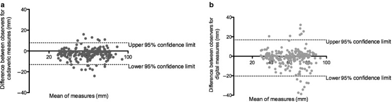
The hip joint is one of the most frequent sites of osteoarthritis. Advances in diagnosis and clinical treatment have progressed dramatically in the last few decades; however, there are limitations associated with the lack of reliable measures for quantifying hip joint morphology. Current diagnostic measures of the hip are performed with pre-determined measures, typically lengths and angles, on 2D radiographic planes. The current measurement techniques do not utilize the inherent 3D nature of CT and MR imaging and do not necessarily quantify the relevant clinical pathologies. A valid and reliable measurement modality that measures the surface geometry of the femoral head is necessary for early diagnosis and treatment of hip disease. The purpose of this study was to establish a method to quantify femoral head morphology using a three-dimensional model. A novel measurement approach was applied to 45 cadaveric femurs (23 right; 22 left; nine female, 17 male) and their digitally reconstructed 3D CT models. The mean difference between the cadaveric and digital measures was -2.04 mm with 95% confidence limits (CI) of 13.67 mm and -17.75 mm, respectively. The digital measurement approach was found to have excellent intraobserver reliability (ICC = 0.99, CI 0.98-0.99) and interobserver reliability (ICC = 0.98, CI 0.93-0.99). This valid and reliable novel digital measurement approach enables quantification of the 3D surface geometry of the femoral head and is able to measure individual variations and potentially detect abnormalities. This method may be used to assist future studies to establish valid diagnostic measurements for femoral head and head-neck junction pathologies.
Keywords: Mimics; cadaveric; hip joint; morphology; morphometrics; reliability; three-dimensional; validity.
© 2014 Anatomical Society.
Figures


References
-
- Audenaert E, Vigneron L, Pattyn C. A method for three-dimensional evaluation and computer aided treatment of femoroacetabular impingement. Comput Aided Surg. 2011;16:143–148. - PubMed
-
- Beaule PE, Zaragoza E, Motamedi K, et al. Three-dimensional computed tomography of the hip in the assessment of femoroacetabular impingement. J Orthop Res. 2005;23:1286–1292. - PubMed
-
- Beck M, Kalhor M, Leunig M, et al. Hip morphology influences the pattern of damage to the acetabular cartilage: femoroacetabular impingement as a cause of early osteoarthritis of the hip. J Bone Joint Surg Br. 2005;87:1012–1018. - PubMed
MeSH terms
LinkOut - more resources
Full Text Sources
Other Literature Sources
