. 2014 Jun 23:5:4234.
doi: 10.1038/ncomms5234.

SUMO1 modification stabilizes CDK6 protein and drives the cell cycle and glioblastoma progression

Affiliations

SUMO1 modification stabilizes CDK6 protein and drives the cell cycle and glioblastoma progression

Anita C Bellail et al. Nat Commun. .

Abstract

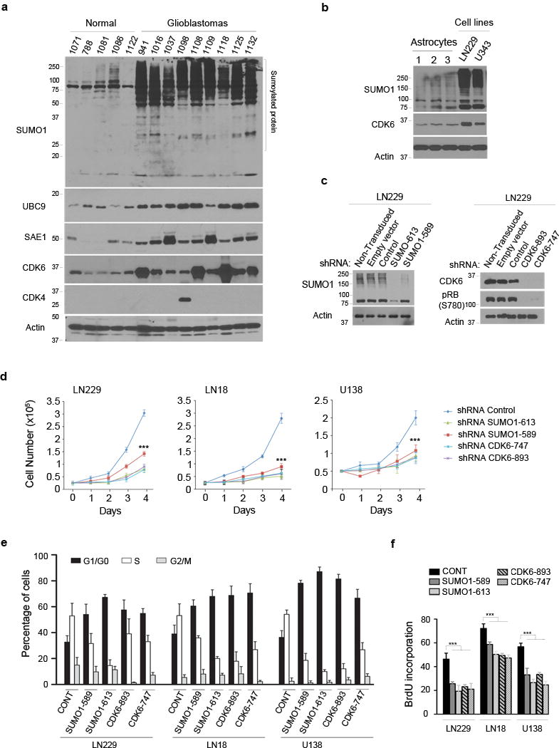
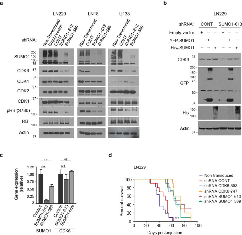
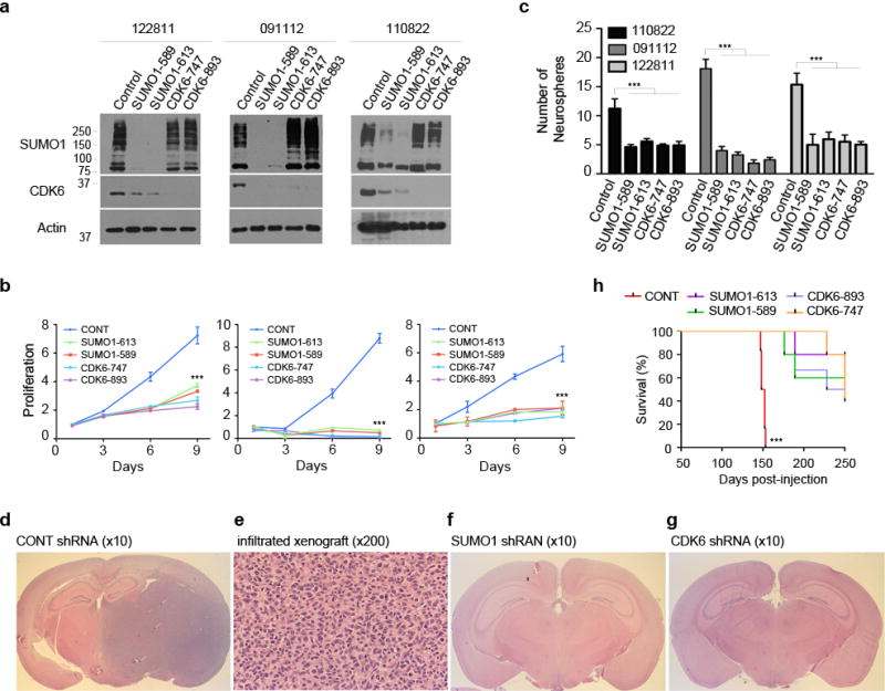
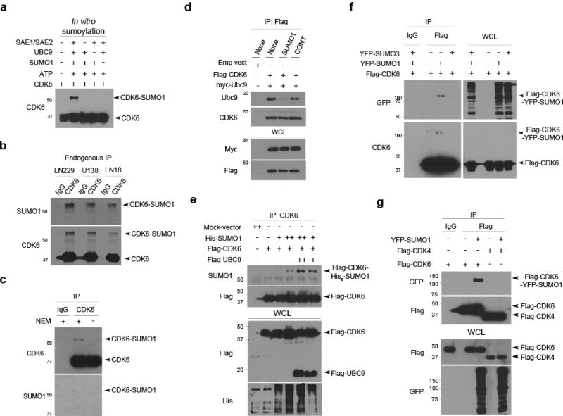
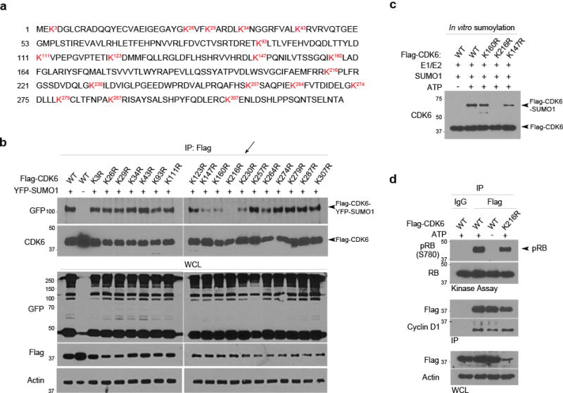
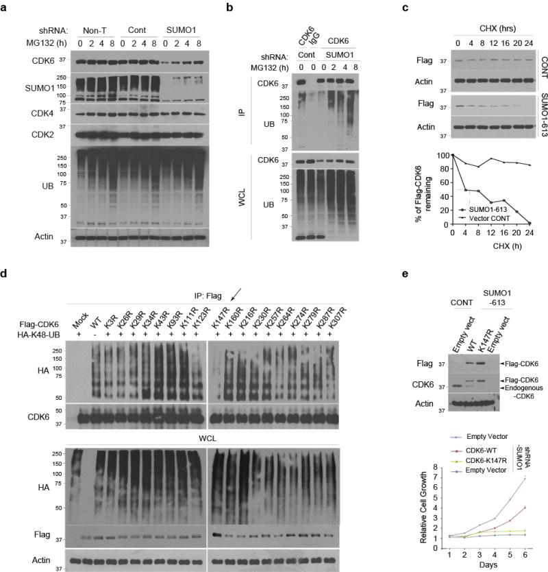
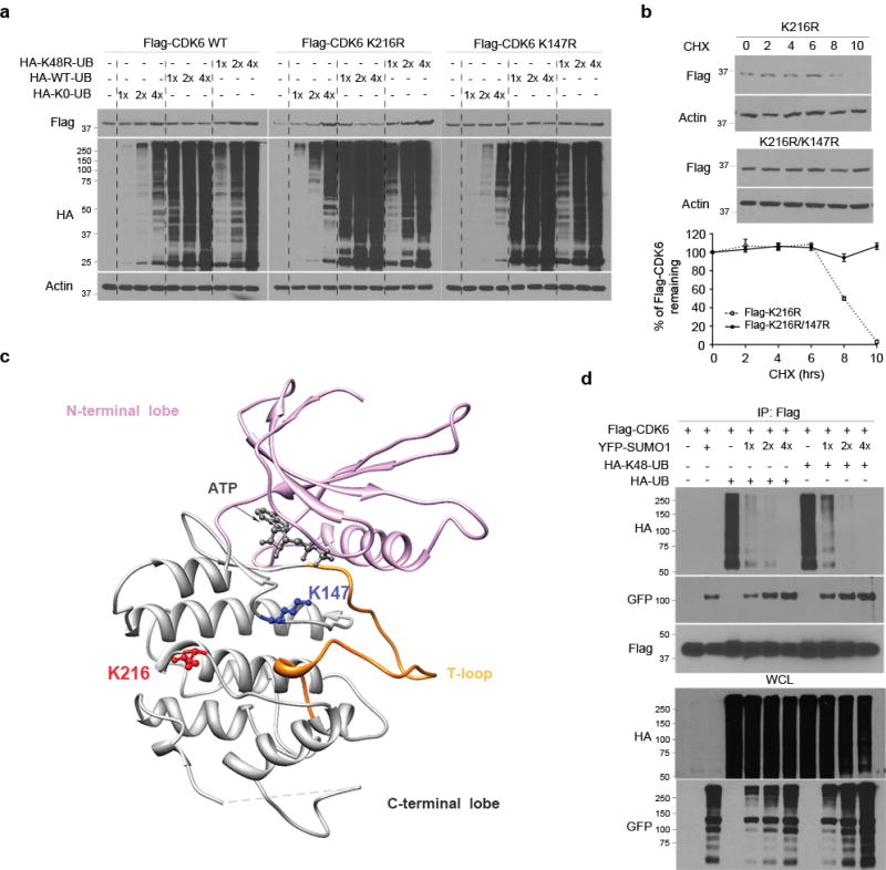
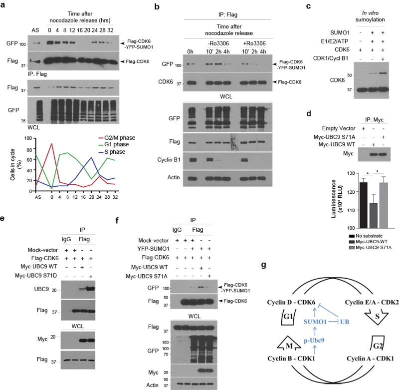
Ubiquitination governs oscillation of cyclin-dependent kinase (CDK) activity through a periodic degradation of cyclins for orderly cell cycle progression; however, the mechanism that maintains the constant CDK protein levels throughout the cell cycle remains unclear. Here we show that CDK6 is modified by small ubiquitin-like modifier-1 (SUMO1) in glioblastoma, and that CDK6 SUMOylation stabilizes the protein and drives the cell cycle for the cancer development and progression. CDK6 is also a substrate of ubiquitin; however, CDK6 SUMOylation at Lys 216 blocks its ubiquitination at Lys 147 and inhibits the ubiquitin-mediated CDK6 degradation. Throughout the cell cycle, CDK1 phosphorylates the SUMO-specific enzyme, ubiquitin-conjugating enzyme9 (UBC9) that in turn mediates CDK6 SUMOylation during mitosis; CDK6 remains SUMOylated in G1 phase and drives the cell cycle through G1/S transition. Thus, SUMO1-CDK6 conjugation constitutes a mechanism of cell cycle control and inhibition of this SUMOylation pathway may provide a strategy for treatment of glioblastoma.


Conflict of interest statement

Competing financial interests: the authors declare no competing financial interests.

Figures

Figure 1
Figure 1. SUMO1 conjugation drives the cell cycle progression through G1/S transition
(a) Normal human brain and glioblastoma tissue samples were examined by western blotting using the indicated antibodies (left). β-actin was used as the protein loading control and the molecular weight were indicated (left). (b) Three human astrocyte samples and two glioblastoma cell lines (top) were tested by western blotting for the expression of SUMO1-conjugated proteins and CDK6. (c) LN229 cells were transduced or not with the empty, scramble control, SUMO1 shRNA as indicated (top) and examined by western blotting using a SUMO1 antibody. (d) Three cell lines transduced with the shRNAs (top) were analyzed for cell growth for 4 days (points: means; bar: SE; n = 6 from two independent experiments; *** P < 0.001; student’s t-test). (e,f) The transduced cell lines were analyzed by flow cytometry for cell cycle (e) and BrdU incorporation (f) (points: means; bar: SD; n = 3 from two independent experiments; *** P < 0.001; student’s t-test).
Figure 2
Figure 2. SUMO1 and CDK6 are required for glioblastoma progression
(a) Three cell lines were transduced or not with the indicated vectors (top) and subjected to western blotting using the antibodies as indicated (left). (b) The SUMO1-613 and control shRNA expressing LN229 cells were transfected either with YFP-SUMO1 or His-SUMO1 and examined by western blotting using CDK6, his and GFP antibody that also recognized YFP. (c) Total RNA isolated from the cells as described above in (a) was examined by real time PCR and the mRNA levels of SUMO1 and CDK6 were normalized to the control β-actin mRNA levels (points: means; bar: SD; n = 6 from two independent experiments; *** P < 0.001; ** P < 0.01; NS: no significance; student’s t-test). (d) Kaplan-Meier curves show the survival of the mice transplanted intracranially with LN229 cells transduced or not with the indicated shRNA vectors (n = 10 for each group; *** P < 0.001; Log rank test).
Figure 3
Figure 3. SUMO1 and CDK6 contributes to the tumorigenesis of neurospheres
(a) Three neurospheres were transduced or not with the SUMO1 and CDK6 shRNAs as indicated (top) and tested by western blotting using SUMO1 and CDK6 antibodies. (b) The transduced neurospheres were examined by cell growth for 9 days (points: means; bar: SD; n = 6 from two independent experiments; *** P < 0.001; student’s t-test). (c) The transduced neurospheres were also examined by sphere formation assay with the numbers of neurospheres as indicated (left) (points: means; bar: SD; n = 6 from three independent experiments; *** P < 0.001; student’s t-test). (d,e) Coronal cerebral section (d) and microscopic section of hematoxylin and eosin (e) reveals the infiltrating glioblastoma in the right side of a mouse brain injected with the neurospheres expressing the control scrambled shRNA. (f,g) The glioblastoma xenograft was not observed in the mouse brain injected with the neurospheres expressing shRNA targeting SUMO1 (f) or CDK6 shRNA (g). (h) Kaplan-Meier curves show the survival of the mice intracranially injected with the neurospheres transduced with the indicated shRNA vectors (n = 6 for each group; *** P < 0.001; Log rank test).
Figure 4
Figure 4. CDK6 is conjugated by SUMO1but not SUMO2/3
(a) In vitro sumoylation assay was performed in a reaction with the elements as indicated (top) and examined by western blotting using a CDK6 antibody with the free and conjugated forms of CDK6 indicated (right). (b) Three cell lines were lysed in a denaturing buffer, immunoprecipitated (IP) with a CDK6 antibody with IgG used as a negative IP control and examined by western blotting using SUMO1 and CDK6 antibody. (c) LN229 cells were lysed in a denaturing buffer supplemented with or without NEM and the endogenous CDK6 protein was immunoprecipitated using a CDK6 antibody with IgG as control and tested examined by western blotting using SUMO1 and another CDK6 antibody. (d) LN229 cells transduced or not with SUMO1 and control shRNA (top) were transfected with Flag-CDK6 and myc-UBC9 (left top). Flag IP and whole cell lysate (WCL) were tested by western blotting for the interaction of Ubc9 and CDK6. (e) LN229 cells were co-transfected with the indicated vectors (top), immunoprecipitated with a CDK6 antibody and examined by western blotting for the presence of Flag-CDK6-His-SUMO1 conjugates (right). (f) LN229 cells were co-transfected with YFP-SUMO3, YFP-SUMO1 and/or Flag-CDK6 and then immunoprecipitated with a Flag antibody. Western blotting using CDK6 and GFP antibody that recognizes YFP detected SUMO1-CDK6 conjugates as indicated (right). (g) Flag immunoprecipitate from YFP-SUMO1, Flag-CDK4 and Flag-CDK6 transfected LN229 cells were examined by western blotting for the presence of Flag-CDK6-YFP-SUMO1 conjugates.
Figure 5
Figure 5. SUMO1 is conjugated to the non-consensus Lys 216 on CDK6 protein
(a) The amino acid sequence of CDK6 reveals 18 lysine residues as indicated (red). (b) Each of the 18 Lys (K) residues was replaced with an arginine (R) through point mutagenesis. Flag-tagged CDK6 wt and K-R mt were transfected with YFP-SUMO1 in LN229 cells. The transfected cells were lysed in a denaturing and diluted in a non-denaturing buffer. The cell lysates were immunoprecipitated with a Flag antibody and examined by western blotting using the indicated antibodies (left), revealing YFP-SUMO1-Flag-CDK6 conjugates in the cells transfected with all except K216R (arrow). (c) In vitro sumoylation assay was carried out in the reaction with the indicated elements (top) and the reaction was analyzed by western blotting using a CDK6 antibody. (d) SUMO1 and control shRNA expressing LN229 cells were transfected with Flag-CDK6 wt and K-R mt. Flag immunoprecipitates (IP) were subjected to kinase assay (top panel) and, together with whole cell lysate (WCL), examined by western blotting (bottom panel).
Figure 6
Figure 6. CDK6 sumoylation inhibits its ubiquitination and degradation
(a) LN229 cells, transduced or not with the indicated shRNAs, were treated with MG132 for the indicated hours (top) and examined by western blotting using the indicated antibodies (left). (b) The transduced cells were immunoprecipitated (IP) by a CDK6 antibody. The IP and whole cell lysate (WCL) were examined by western blotting using UB and another CDK6 antibody. (c) The half-life of Flag-CDK6 protein was evaluated by western blotting (top) and quantified against the control β-actin protein in SUMO1 and control shRNA expressing cells (bottom). (d) SUMO1 shRNA expressing LN229 cells were transfected with Flag-CDK6 wt and K-R mt with HA-K48-polyubiquitin (HA-K48-UB). Flag immunoprecipitates were examined by western blotting using HA and CDK6 antibodies. (e) The SUMO1-613 shRNA expressing LN229 cells were transfected with Flag-CDK6 wt, K147R and empty vector and analyzed by western blotting (top) and cell growth assay (bottom) (points: means; bar: SD; n = 6 from two independent experiments; *** P < 0.001; student’s t-test).
Figure 7
Figure 7. Sumoylation and ubiquitination regulate CDK6 degradation
(a) Flag-CDK6 wt, K216R and K147R mt were transfected in LN229 cells with HA-K48R polyubiquitin (HA-48-UB), HA-ubiquitin (HA-UB) and/or non-conjugated ubiquitin mt (K0-UB) in the concentrations as indicated (top). Flag-CDK6 and HA-ubiquitin were detected by western blotting using Flag and HA antibodies. (b) The half-life of Flag-CDK6 K216R and K147R mt were evaluated by western blotting (top) and quantified against the control β-actin in SUMO1 and control shRNA expressing cells (bottom) (points: means; bar: SD; n = 6 from two independent experiments). (d) The CDK6 protein structure, generated at http://www.rcsb.org (PDB 2EUF), shows the kinase active site with the ATP binding side chains in the deep cleft between N-terminal and C-terminal lobe from which T-loop rises. K216 of the C-terminal is close to K147 near the kinase active site. (d) LN229 cells were transfected with Flag-CDK6, HA-K48-UB, HA-UB and YFP-SUMO1. Flag immunoprecipitates (IP) and whole cell lysate (WCL) were tested by western blotting using the indicated antibodies (left).
Figure 8
Figure 8. CDK1 activity phosphorylates UBC9 for CDK6 sumoylation
(a) YFP-SUMO1 and Flag-CDK6 expressing LN229 cells were immunoprecipitated in the hours after released from nocodazole synchronization to the cell cycle as monitored by flow cytometry (low panel). Flag immunoprecipitates (IP) and whole cell lysate (WCL) were analyzed by western blotting with asynchronized (AS) cells used as controls (top panel). (b) LN229 cells were co-transfected with YFP-SUMO1 and Flag-CDK6, treated with nocodazole for 20 hrs and then treated or not with RO-3306 for the indicated hrs (top). Flag IP and WCL were analyzed by western blotting using the indicated antibodies (left). (c) In vitro sumoylation of CDK6 in the absence and presence of the cyclin B1-CDK1 complex (CDK1/Cycl B1). (d) HEK293 cells were expressed with the tagged proteins, immunoprecipitated and analyzed by western blotting (top) and CDK1 kinase assay (bottom). (e) Flag-CDK6 was transfected with Myc-UBC9 wt or S71D mt in LN229 cells, immunoprecipitated using Flag antibodies and tested by western blotting for UBC9-CDK6 interaction. (f) LN229 cells were transfected with Myc-UBC9 WT and S71A mt together with Flag-CDK6 and/or YFP-SUMO1, immunoprecipitated and examined by western blotting. (g) The molecular model shows that CDK1 phosphorylates UBC9 and thus enhances SUMO1-CDK6 conjugation from G2/M to G1 phase, driving the cell cycle through G1/S transition.

References

    1. Hay RT. SUMO: a history of modification. Mol Cell. 2005;18:1–12. - PubMed
    1. Geiss-Friedlander R, Melchior F. Concepts in sumoylation: a decade on. Nat Rev Mol Cell Biol. 2007;8:947–956. - PubMed
    1. Gareau JR, Lima CD. The SUMO pathway: emerging mechanisms that shape specificity, conjugation and recognition. Nat Rev Mol Cell Biol. 2010;11:861–871. - PMC - PubMed
    1. Bhoj VG, Chen ZJ. Ubiquitylation in innate and adaptive immunity. Nature. 2009;458:430–437. - PubMed
    1. Bernier-Villamor V, Sampson DA, Matunis MJ, Lima CD. Structural basis for E2-mediated SUMO conjugation revealed by a complex between ubiquitin-conjugating enzyme Ubc9 and RanGAP1. Cell. 2002;108:345–356. - PubMed

Publication types

MeSH terms