. 2014 Aug;76(2):279-95.
doi: 10.1002/ana.24204. Epub 2014 Jul 2.

Functional anatomy of subthalamic nucleus stimulation in Parkinson disease

Affiliations

Functional anatomy of subthalamic nucleus stimulation in Parkinson disease

Sarah A Eisenstein et al. Ann Neurol. 2014 Aug.

Abstract

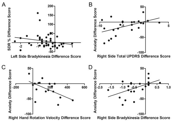
Objective: We developed a novel method to map behavioral effects of deep brain stimulation (DBS) across a 3-dimensional brain region and to assign statistical significance after stringent type I error correction. This method was applied to behavioral changes in Parkinson disease (PD) induced by subthalamic nucleus (STN) DBS to determine whether these responses depended on anatomical location of DBS.

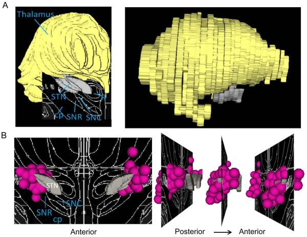
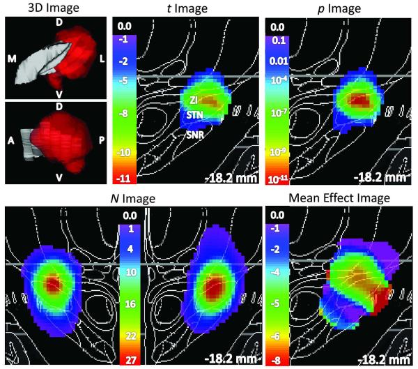
Methods: Fifty-one PD participants with STN DBS were evaluated off medication, with DBS off and during unilateral STN DBS with clinically optimized settings. Dependent variables included DBS-induced changes in Unified Parkinson Disease Rating Scale (UPDRS) subscores, kinematic measures of bradykinesia and rigidity, working memory, response inhibition, mood, anxiety, and akathisia. Weighted t tests at each voxel produced p images showing where DBS most significantly affected each dependent variable based on outcomes of participants with nearby DBS. Finally, a permutation test computed the probability that this p image indicated significantly different responses based on stimulation site.

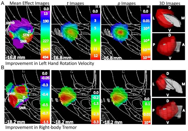
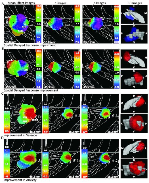
Results: Most motor variables improved with DBS anywhere in the STN region, but several motor, cognitive, and affective responses significantly depended on precise location stimulated, with peak p values in superior STN/zona incerta (quantified bradykinesia), dorsal STN (mood, anxiety), and inferior STN/substantia nigra (UPDRS tremor, working memory).

Interpretation: Our method identified DBS-induced behavioral changes that depended significantly on DBS site. These results do not support complete functional segregation within STN, because movement improved with DBS throughout, and mood improved with dorsal STN DBS. Rather, findings support functional convergence of motor, cognitive, and limbic information in STN.


Figures

Figure 1
Figure 1. 3D models of the STN, thalamus, and DBS electrode contacts
A.) Brain regions are overlaid on the Mai atlas (18.2 mm posterior to AC) in coronal (left) and sagittal (right) views. B.) The 3D distribution of clinical STN DBS electrode contacts are presented coronally (left) and sagittally (right), overlaid on the structures from the Mai atlas at 16.8 mm posterior to AC. Most clinical DBS contacts fall in the STN. STN, subthalamic nucleus; cp, cerebellar peduncle; SNR, substantia nigra pars reticulata; SNC, substantia nigra pars compacta; R, red nucleus. Gray = STN; pink spheres = approximate contact location, 1.5 mm radius for display.
Figure 2
Figure 2. STN DBS-induced improvement in UPDRS III motor scores did not differ significantly by DBS site
The left panel shows 3D views of the left STN in gray, with color indicating voxels for which p < 0.05 by weighted t test (upper image: viewed from a point anterior to STN; lower image: viewed from a point lateral to STN). Statistical images in color, laid over coronal slices from the atlas, are shown in the top center panel (t image, thresholded at N ≥ 6), top right panel (p image, thresholded at p < 0.05), bottom left panel (N image) and bottom right panel (weighted mean effect image, thresholded at N ≥ 6). Notably, although the p image includes values as low as 10−11, the improvement did not differ significantly by contact location (p = 0.12 by permutation test; see Results). D, dorsal; V, ventral; A, anterior; P, posterior; M, medial; L, lateral. ZI, zona incerta; SNR, substantia nigra.
Figure 3
Figure 3. STN DBS-induced improvements in hand rotation velocity and tremor significantly differed by DBS site
A.) Left hand rotation velocity: mean effect, t, and p images are shown left to right, with the p image thresholded at p ≤ 0.05, on a Mai atlas section close to the peak p value in black and white at the indicated distance from the plane through the anterior commissure and normal to the bicommissural line; the rightmost panels show 3D representation of voxels for which p ≤ 0.05 in color, with the STN in gray (top left panel: viewed from the front, bottom left panel: viewed from the side). B.) Right-side tremor: same conventions as in 3A. Images are horizontally flipped for clarity. Color scales indicate weighted mean, t, and p values. D, dorsal; V, ventral; A, anterior; P, posterior; M, medial; L, lateral; ZI, zona incerta; STN, subthalamic nucleus; SNR, substantia nigra pars reticulata; comb, comb system (SNR fragments in white matter)
Figure 4
Figure 4. STN DBS-induced alterations in non-motor outcomes significantly differed by DBS site
A.) Impairment in SDR performance. B.) Improvement in SDR performance. Same conventions as in Fig 3 except that weighted mean, t, and p values are shown on all 2D panels for both impairment (aquamarine-violet) and improvement (green-red). C.) Improvement in valence: same conventions as in Fig 3. D.) Improvement in anxiety: same conventions as in Fig 3.
Figure 5
Figure 5. STN DBS-induced changes in cognition and anxiety are related to changes in motor function
A.) Right-brain STN DBS-induced percent change in SDR error was negatively related to right-brain STN DBS-induced improvement in UPDRS subscore bradykinesia. Left-brain STN DBS-induced decreases in anxiety were associated with left-brain STN DBS-induced improvements in B.) right side total UPDRS scores, C.) right hand rotation velocity and D.) right side UPDRS subscore bradykinesia. Except for hand rotation velocity, decreasing difference scores signify improvement in that measure.

Similar articles

Cited by

References

    1. Foltynie T, Hariz MI. Surgical management of Parkinson’s disease. Expert Rev Neurother. 2010;10:903–914. - PubMed
    1. Funkiewiez A, Ardouin C, Caputo E, et al. Long term effects of bilateral subthalamic nucleus stimulation on cognitive function, mood, and behaviour in Parkinson’s disease. J Neurol Neurosurg Psychiatry. 2004;75:834–839. - PMC - PubMed
    1. Smeding HM, Speelman JD, Koning-Haanstra M, et al. Neuropsychological effects of bilateral STN stimulation in Parkinson disease: A controlled study. Neurology. 2006;66:1830–1836. - PubMed
    1. Parent A, Hazrati LN. Functional anatomy of the basal ganglia I. The cortico-basal ganglia-thalamo-cortical loop. Brain Res Brain Res Rev. 1995a;20:91–127. - PubMed
    1. Parent A, Hazrati LN. Functional anatomy of the basal ganglia. II. The place of subthalamic nucleus and external pallidum in basal ganglia circuitry. Brain Res Brain Res Rev. 1995b;20:128–154. - PubMed

Publication types