

The Neurospora crassa CPS-1 polysaccharide synthase functions in cell wall biosynthesis
- PMID: 24953997
- PMCID: PMC4115004
- DOI: 10.1016/j.fgb.2014.05.009
The Neurospora crassa CPS-1 polysaccharide synthase functions in cell wall biosynthesis
Abstract
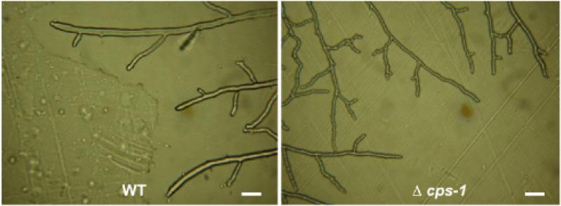
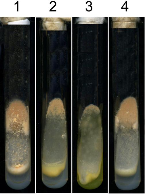
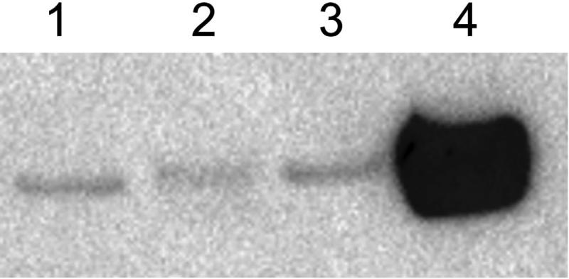
The Neurospora crassa cps-1 gene encodes a polysaccharide synthase with homology to the Cryptococcus neoformans hyaluronic acid synthase Cps1p. Homologs of the cps-1 gene are found in the genomes of many fungi. Loss of CPS-1 results in a cell wall defect that affects all stages of the N. crassa life cycle, including vegetative growth, protoperithecia (female mating structure) development, and conidia (asexual spore) development. The cell wall of cps-1 deletion mutants is sensitive to cell wall perturbation reagents. Our results demonstrate that CPS-1 is required for the incorporation of cell wall proteins into the cell wall and plays a critical role in cell wall biogenesis. We found that the N. crassa cell wall is devoid of hyaluronic acid, and conclude that the polysaccharide produced by the CPS-1 is not hyaluronic acid.
Keywords: Cell wall biogenesis; Cell wall glucan; Cell wall polysaccharide; Fungal cell wall; Neurospora crassa; Polysaccharide synthase.
Copyright © 2014 Elsevier Inc. All rights reserved.
Figures


Similar articles
-
Genetic and biochemical characterization of the GH72 family of cell wall transglycosylases in Neurospora crassa.Fungal Genet Biol. 2017 Apr;101:46-54. doi: 10.1016/j.fgb.2017.03.002. Epub 2017 Mar 8. Fungal Genet Biol. 2017. PMID: 28285007 Free PMC article.
-
Neurospora crassa 1,3-α-glucan synthase, AGS-1, is required for cell wall biosynthesis during macroconidia development.Microbiology (Reading). 2014 Aug;160(Pt 8):1618-1627. doi: 10.1099/mic.0.080002-0. Epub 2014 May 20. Microbiology (Reading). 2014. PMID: 24847001 Free PMC article.
-
Mannosyltransferase is required for cell wall biosynthesis, morphology and control of asexual development in Neurospora crassa.Mycologia. 2005 Jul-Aug;97(4):872-9. doi: 10.3852/mycologia.97.4.872. Mycologia. 2005. PMID: 16457356
-
Recent advances in septum biogenesis in Neurospora crassa.Adv Genet. 2013;83:99-134. doi: 10.1016/B978-0-12-407675-4.00003-1. Adv Genet. 2013. PMID: 23890213 Review.
-
Biochemical genetics of Neurospora crassa conidial germination.Bacteriol Rev. 1976 Mar;40(1):1-41. doi: 10.1128/br.40.1.1-41.1976. Bacteriol Rev. 1976. PMID: 5072 Free PMC article. Review. No abstract available.
Cited by
-
The Glycosylphosphatidylinositol-Anchored DFG Family Is Essential for the Insertion of Galactomannan into the β-(1,3)-Glucan-Chitin Core of the Cell Wall of Aspergillus fumigatus.mSphere. 2019 Jul 31;4(4):e00397-19. doi: 10.1128/mSphere.00397-19. mSphere. 2019. PMID: 31366710 Free PMC article.
-
Characterization of the Neurospora crassa GH72 family of Laminarin/Lichenin transferases and their roles in cell wall biogenesis.Cell Surf. 2025 Jan 3;13:100140. doi: 10.1016/j.tcsw.2024.100140. eCollection 2025 Jun. Cell Surf. 2025. PMID: 39866863 Free PMC article.
-
cpsA regulates mycotoxin production, morphogenesis and cell wall biosynthesis in the fungus Aspergillus nidulans.Mol Microbiol. 2017 Jul;105(1):1-24. doi: 10.1111/mmi.13682. Epub 2017 Apr 24. Mol Microbiol. 2017. PMID: 28370587 Free PMC article.
-
Genetic and biochemical characterization of the GH72 family of cell wall transglycosylases in Neurospora crassa.Fungal Genet Biol. 2017 Apr;101:46-54. doi: 10.1016/j.fgb.2017.03.002. Epub 2017 Mar 8. Fungal Genet Biol. 2017. PMID: 28285007 Free PMC article.
-
Characterization of the putative polysaccharide synthase CpsA and its effects on the virulence of the human pathogen Aspergillus fumigatus.PLoS One. 2019 Apr 26;14(4):e0216092. doi: 10.1371/journal.pone.0216092. eCollection 2019. PLoS One. 2019. PMID: 31026268 Free PMC article.
References
-
- Bowman SM, Piwowar A, Al Dabbous M, Vierula J, Free SJ. Mutational analysis of the glycosylphosphatidylinositol (GPI) anchor pathway demonstrates that GPI-anchored proteins are required for cell wall biogenesis and normal hyphal growth in Neurospora crassa. Eukaryot Cell. 2006;5:587–600. - PMC - PubMed
-
- Cabib E, Blanco N, Grau C, Rodriguez-Pena JM, Arroyo J. Crh1p and Crh2p are required for the cross-linking of chitin to beta(1-6)glucan in the Saccharomyces cerevisiae cell wall. Mol Microbiol. 2007;63:921–35. - PubMed
Publication types
MeSH terms
Substances
Grants and funding
LinkOut - more resources
Full Text Sources
Other Literature Sources
