Review
. 2015 Feb;1849(2):142-51.
doi: 10.1016/j.bbagrm.2014.06.005. Epub 2014 Jun 17.

Estrogen receptor signaling during vertebrate development

Affiliations
Review

Estrogen receptor signaling during vertebrate development

Maria Bondesson et al. Biochim Biophys Acta. 2015 Feb.

Abstract

Estrogen receptors are expressed and their cognate ligands produced in all vertebrates, indicative of important and conserved functions. Through evolution estrogen has been involved in controlling reproduction, affecting both the development of reproductive organs and reproductive behavior. This review broadly describes the synthesis of estrogens and the expression patterns of aromatase and the estrogen receptors, in relation to estrogen functions in the developing fetus and child. We focus on the role of estrogens for the development of reproductive tissues, as well as non-reproductive effects on the developing brain. We collate data from human, rodent, bird and fish studies and highlight common and species-specific effects of estrogen signaling on fetal development. Morphological malformations originating from perturbed estrogen signaling in estrogen receptor and aromatase knockout mice are discussed, as well as the clinical manifestations of rare estrogen receptor alpha and aromatase gene mutations in humans. This article is part of a Special Issue entitled: Nuclear receptors in animal development.

Keywords: Aromatase; Estrogen; Estrogen receptor; Reproductive development; Sex differentiation; Vertebrate development.


Figures

Figure 1
Figure 1
Schematic representation of selected steroidogenic pathways for synthesis of estrogens. Different pathways are present in different tissues. Estrogens are shown in blue, and steroidogenic enzymes in red. DHEAS=Dehydroepiandrosterone sulfate; 5-Diol=5-Androstene-3β, 17β-diol; DHEA=Dehydroepiandrosterone; 5α-Dione=5-alpha-androstane-3,17-dione; ADT=Androsterone, epiADT=epi-Androsterone; DHT=Dihydrotestosteron, 3β-Adiol=5α-androstane-3β, 17β-diol; E1=Estrone; E1S=Estrone sulfate; E2=17β-estradiol; E2S=17β-eEtradiol sulfate; 3αAdiol=5α-androstane-3α,17β-diol; E3=Estriol; E4=Estetrol; Cyp=Cytochrome P450; and HSD=Hydroxysteroid dehydrogenase. Modified from (Labrie et al., 2005) and Warmerdam (Warmerdam et al., 2008).
Figure 2
Figure 2
Schematic representation of expression of ERs and aromatase during fetal development in mouse, and in the human fetus based on qPCR and immunohistochemistry data. The mouse RNA expression data was extracted from NCBI’s Unigene (www.ncbi.nlm.nih.gov/UniGene). The human fetal data is described in publications as referenced in the main text. For the mouse fetus, the Unigene database does not report any expression of aromatase, whereas publications do (Greco and Payne, 1994; Harada and Yamada, 1992). This conflicting information has been marked by +/−.
Figure 3
Figure 3
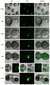
GFP expression visualizing estrogen activity in zebrafish embryos from 6 hpf to 6 dpf. First row: Maternal effect of Tg(5xERE:GFP) transgenic fish at 6 hpf in the absence of E2. Scale bars, 500 μm. Second to seventh rows: E2-induced GFP expression in Tg(5xERE:GFP) transgenic fish during development. Zebrafish larvae were treated with 1 μM E2 (in 0.1% DMSO) or vehicle alone (control, 0.1% DMSO) from 3 hpf and imaged at 6 hpf, 1 dpf, 2 dpf, 3 dpf, 4 dpf, 5 dpf and 6 dpf. First column, bright-field images; second column, corresponding GFP fluorescence images; third column, overlay of bright-field and GFP images. 1dpf, lateral view; 2–4 dpf, dorsal view; 5 dpf, left panel, dorsal, and right panel, ventral view; anterior to the left. Scale bars, 100 μm. Adapted from (Hao et al., 2013).

References

    1. Aidoo-Gyamfi K, Cartledge T, Shah K, Ahmed S. Estrone sulfatase and its inhibitors. Anti-cancer agents in medicinal chemistry. 2009;9:599–612. - PubMed
    1. Al Sweidi S, Sanchez MG, Bourque M, Morissette M, Dluzen D, Di Paolo T. Oestrogen receptors and signalling pathways: implications for neuroprotective effects of sex steroids in Parkinson’s disease. Journal of neuroendocrinology. 2012;24:48–61. - PubMed
    1. Albalat R, Brunet F, Laudet V, Schubert M. Evolution of retinoid and steroid signaling: vertebrate diversification from an amphioxus perspective. Genome biology and evolution. 2011;3:985–1005. - PMC - PubMed
    1. Albrecht ED, Lane MV, Marshall GR, Merchenthaler I, Simorangkir DR, Pohl CR, Plant TM, Pepe GJ. Estrogen promotes germ cell and seminiferous tubule development in the baboon fetal testis. Biology of reproduction. 2009;81:406–414. - PMC - PubMed
    1. Aquila S, Sisci D, Gentile M, Middea E, Siciliano L, Ando S. Human ejaculated spermatozoa contain active P450 aromatase. The Journal of clinical endocrinology and metabolism. 2002;87:3385–3390. - PubMed

Publication types

LinkOut - more resources