. 2014 Nov;22(11):1910-22.
doi: 10.1038/mt.2014.117. Epub 2014 Jun 23.

Targeted inhibition of prostate cancer metastases with an RNA aptamer to prostate-specific membrane antigen

Affiliations

Targeted inhibition of prostate cancer metastases with an RNA aptamer to prostate-specific membrane antigen

Justin P Dassie et al. Mol Ther. 2014 Nov.

Abstract

Cell-targeted therapies (smart drugs), which selectively control cancer cell progression with limited toxicity to normal cells, have been developed to effectively treat some cancers. However, many cancers such as metastatic prostate cancer (PC) have yet to be treated with current smart drug technology. Here, we describe the thorough preclinical characterization of an RNA aptamer (A9g) that functions as a smart drug for PC by inhibiting the enzymatic activity of prostate-specific membrane antigen (PSMA). Treatment of PC cells with A9g results in reduced cell migration/invasion in culture and metastatic disease in vivo. Importantly, A9g is safe in vivo and is not immunogenic in human cells. Pharmacokinetic and biodistribution studies in mice confirm target specificity and absence of non-specific on/off-target effects. In conclusion, these studies provide new and important insights into the role of PSMA in driving carcinogenesis and demonstrate critical endpoints for the translation of a novel RNA smart drug for advanced stage PC.


Figures

Figure 1
Figure 1
PSMA expression drives cancer cell proliferation, migration and invasion. (a) Proliferation of PC-3(PSMA-) and PC-3(PSMA+) cells grown in culture determined by MTS assay. (Inset) Fold increase in the rate of proliferation from 24 to 48 hours (b) (Left panel) Migration of PC-3(PSMA-) and PC-3(PSMA+) cells determined by scratch-wound assay. (Right panel) Migration of CT26(PSMA) and CT26(PSMA+) cells determined by scratch-wound assay. *P < 0.05, **P < 0.001 (c) Invasion of CT26 and PC3 cells, expressing varying amounts of PSMA on the cell surface, was determined by Matrigel invasion assay. () = no PSMA expression, (+) = low PSMA expression, (++) = high PSMA expression as determined by flow cytometry analysis MTS, 3-(4 5-dimethylthiazol-2-yl)-5-(3-carboxymethoxyphenyl)-2-(4-sulfophenyl)-2h-tetrazolium.
Figure 2
Figure 2
Functional characterization of PSMA RNA aptamers. (a) Predicted secondary structures of PSMA-specific aptamers A10-3.218 and A9g, as well as, control non-binding aptamer (A9g.6). Arrows indicate the nucleotide that was mutated in A9g to generate the non-binding, control aptamer A9g.6. Structural predictions were generated using RNA structure version 5.03. (b) Effect of PSMA aptamers on PSMA enzymatic activity determined by NAALADase Assay. (c) Effect of A9g on PSMA-mediated cell proliferation determined by MTS Assay. Doxorubicin: antiproliferative chemotherapeutic drug; 2-PMPA: small molecule inhibitor of PSMA enzymatic activity. (d) Effect of A9g on PSMA-mediated cell migration determined by transwell migration assay. (e) Effect of A9g on PSMA-mediated cell invasion determined by Matrigel invasion assay MTS, 3-(4 5-dimethylthiazol-2-yl)-5-(3-carboxymethoxyphenyl)-2-(4-sulfophenyl)-2h-tetrazolium.
Figure 3
Figure 3
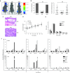
In vivo efficacy and safety of A9g PSMA aptamer. (a) Representative images of vehicle (n = 10), A9g (n = 18) and A9g.6 (n = 16) treated SCID mice following intra-cardiac injection of luciferase (Luc+) expressing PSMA+ prostate cancer cells, 22Rv1(1.7). Images were acquired on the AMI 1000 Instrument (Spectral Instruments Imaging). (b) (Left panel) Percentage of mice with metastases from the three treatment groups (vehicle, A9g or A9g.6) 4 weeks after intra-cardiac injection of 22rv1(1.7) cells (*P < 0.0001, Fisher's exact test comparing A9g-treated group to either vehicle or A9g.6-treated groups). (Right panel) Number of bioluminescent (Luc+) foci (metastatic foci) per mouse, per group was quantified. (**P < 0.001, Student t-test comparing A9g-treated group to either vehicle or A9g.6-treated groups) (c) Representative histological section of bone with disseminated disease. Hematoxylin and Eosin (H&E) staining of bone section at 100× and 400× shown * = indicates bone metastases. (d) Effect of A9g aptamer on mouse weight during course of treatment. Normalized weight: weights normalized to pretreatment values. (e) Effect of A9g on blood cells after a complete treatment course. CBC is reported for white blood cells (WBC), red blood cells (RBC), platelets (PLT), neutrophils (NEU), lymphocytes (LYM), and monocytes (MON). (f) Assessment of potential immune stimulatory effect of A9g in immune-competent mice. Poly I:C: positive control for immune stimulation. Spleens (top panels) and livers (bottom panels) of treated mice were collected at the indicated time points and processed for total RNA. Expression levels of several immune responsive genes were determined by RT-qPCR. OAS-1: 2′-5′ oligoadenylate synthetase 1A, IFN-β: interferon beta 1, IFN-γ: interferon gamma, IL-6: interleukin 6, and IFIT1: interferon-induced protein with tetratricopeptide repeats 1.
Figure 4
Figure 4
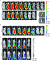
PK and biodistribution of PSMA aptamer, A9g. (a) Near NIR labeled aptamers NIR-A9g and NIRA9g.6 were injected tail vein in mice bearing PSMA+ tumors (implanted subcutaneously) and tracked over time with the IVIS 200 imaging system (PerkinElmer). Ex-vivo: tumors were excised at day 4 (96 hours) post-injection. (b) PK and biodistribution of NIR-A9g in a control, non-tumor-bearing mouse. (c) Specific targeting of PSMA positive tumor by A9g. NIR-A9g (top row) or NIR-A9g.6 (bottom row) were injected tail vein in SCID mice bearing subcutaneous PSMA+ (right flank) or PSMA (left flank) prostate cancer tumors. Images were acquired with the IVIS 200 imaging system (PerkinElmer) at the indicated time points following injection of the labeled RNAs.
Figure 5
Figure 5
Stability and safety assessment of A9g aptamer in human serum and cells. (a) Effect of chemical modifications on stability of A9g aptamer in human serum. (Left panel) Denaturing PAGE gel of RNA aptamers following incubation with 100% human serum over a 1-week period. (Right panel) Band intensity was quantified using Image J version 1.47 and plotted relative to input/time 0 RNA. A9g = All pyrimidines modified with 2′-fluoro chemistry, A9g + 3 = All pyrimidines modified with 2′-fluoro chemistry and three purines modified with 2′-O methyl chemistry; A9g + 21 = All pyrimidines modified with 2′-fluoro chemistry and all 21 purines modified with 2′-O methyl chemistry. (b) Functional characterization of chemically modified A9g aptamers using NAALADAse Assay. Percentage of PSMA enzymatic activity is reported. (c) Assessment of potential toxicity of A9g in human peripheral blood mononuclear cells (hPBMC) from healthy adult volunteers. Staurosporine: positive control for apoptosis; Poly I:C: positive control for immune stimulation. Vehicle: Binding Buffer or a non-binding, control aptamer (A9g.6) for the indicated time points. (Left panel) Apoptosis assessed by caspase 3/7 activation. (Right panel) Cytotoxicity assessed by lactate dehydrogenase activity. (d) Assessment of potential immune stimulatory effect of A9g in hPBMCs as in Figure 3f for above.

Comment in

Similar articles

Cited by

References

    1. Cereda V, Formica V, Massimiani G, Tosetto L, Roselli M. Targeting metastatic castration-resistant prostate cancer: mechanisms of progression and novel early therapeutic approaches. Expert Opin Investig Drugs. 2014;23:469–487. - PubMed
    1. Pal SK, Twardowski P, Sartor O. Critical appraisal of cabazitaxel in the management of advanced prostate cancer. Clin Interv Aging. 2010;5:395–402. - PMC - PubMed
    1. Shah N, Dizon DS. New-generation platinum agents for solid tumors. Future Oncol. 2009;5:33–42. - PubMed
    1. Ghosh A, Heston WD. Tumor target prostate specific membrane antigen (PSMA) and its regulation in prostate cancer. J Cell Biochem. 2004;91:528–539. - PubMed
    1. Israeli RS, Powell CT, Corr JG, Fair WR, Heston WD. Expression of the prostate-specific membrane antigen. Cancer Res. 1994;54:1807–1811. - PubMed

Publication types