

Comparison of extracapsular pseudotumors seen in magnetic resonance imaging and in revision surgery of 167 failed metal-on-metal hip replacements
- PMID: 24954485
- PMCID: PMC4164864
- DOI: 10.3109/17453674.2014.934189
Comparison of extracapsular pseudotumors seen in magnetic resonance imaging and in revision surgery of 167 failed metal-on-metal hip replacements
Abstract
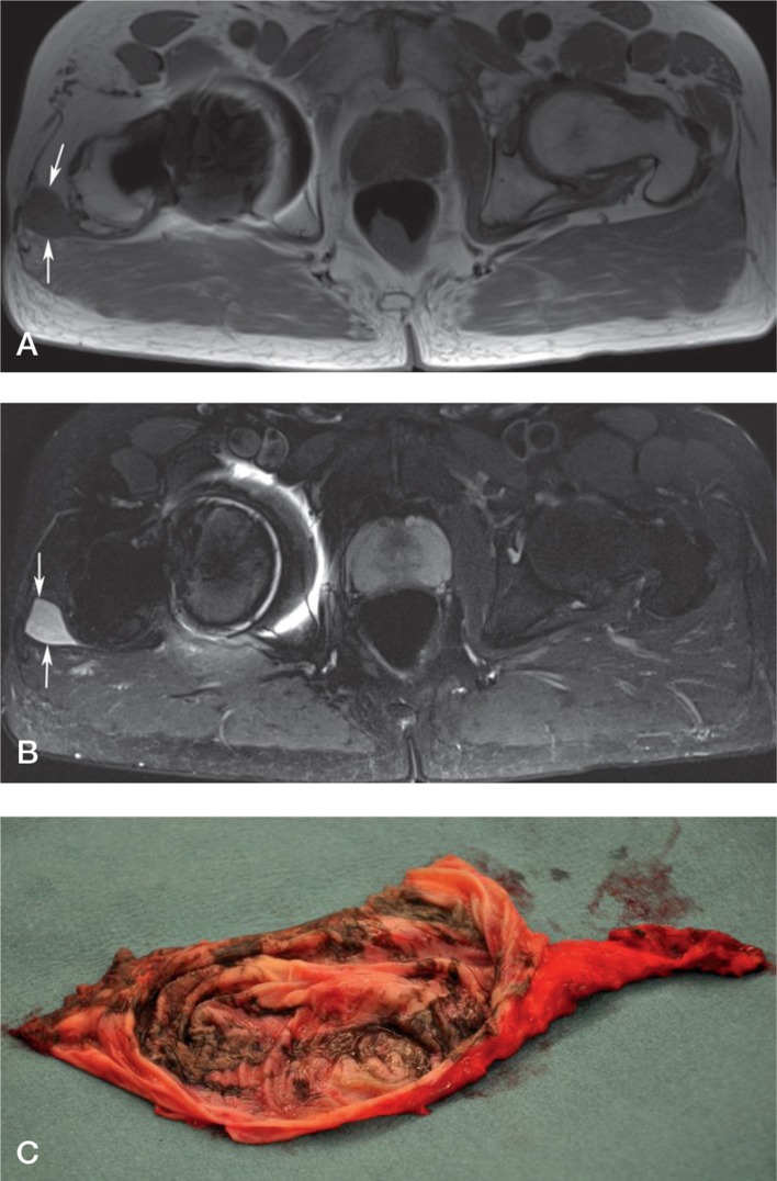
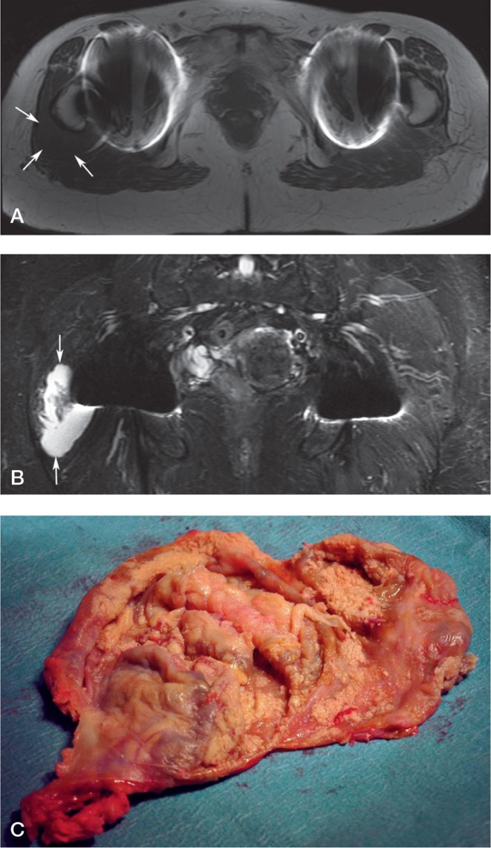
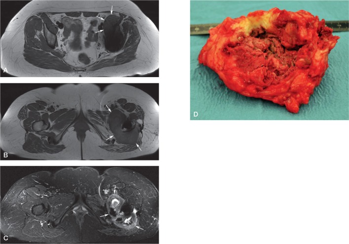
Background and purpose: Magnetic resonance imaging (MRI) is important for detecting extracapsular pseudotumors, but there is little information on the accuracy of MRI and appropriate intervals for repeated imaging. We evaluated the sensitivity and specificity of MRI for detecting pseudotumors in 155 patients (167 hips) with metal-on-metal (MoM) hip arthroplasties that failed due to adverse reactions to metal debris (ARMD).
Methods: Preoperative MRIs were performed with two 1.5 T MRI scanners and graded by a senior musculoskeletal radiologist using a previously described MRI pseudotumor grading system. Revision findings were retrieved from surgical notes, and pseudotumors were retrospectively graded as fluid-filled, mixed-type, or solid.
Results: The sensitivity of MRI was 71% and the specificity was 87% for detecting extracapsular pseudotumors. The sensitivity was 88% (95% CI: 70-96) when MRI was performed less than 3 months before the revision surgery. Interestingly, when the time that elapsed between MRI and revision was more than 1 year, the sensitivity calculated was only 29% (95% CI: 14-56). Comparison between MRI and revision classifications gave moderate agreement (Cohen's kappa = 0.4).
Interpretation: A recent MRI predicts the presence of a pseudotumor well, but there is more discrepancy when the MRI examination is over a year old, most likely due to the formation of new pseudotumors. 1 year could be a justifiable limit for considering a new MRI if development of ARMD is suspected. MRI images over a year old should not be used in decision making or in planning of revision surgery for MoM hips.
Figures
References
-
- Almousa SA, Greidanus NV, Masri BA, Duncan CP, Garbuz DS. The natural history of inflammatory pseudotumors in asymptomatic patient... . Clin Orthop. 2013;471(12):3814–21. - PMC - PubMed
-
- Anderson H, Toms AP, Cahir JG, Goodwin RW, Wimhurst J, Nolan JF. Grading the severity of soft tissue changes associated with metal-on-met... . Skeletal Radiol. 2011;40(3):303–7. - PubMed
-
- Bozic KJ, Kurtz S, Lau E, Ong K, Chiu V, Vail TP, Rubash HE, Berry DJ. The epidemiology of bearing surface usage in total hip arthroplasty in t... . J Bone Joint Surg (Am) 2009;91(7):1614–20. - PubMed
-
- Chang EY, McAnally JL, Van Horne JR, Statum S, Wolfson T, Gamst A, Chung CB. Metal-on-metal total hip arthroplasty: Do symptoms correlate with MR ima... . Radiology. 2012;265(3):848–57. - PubMed
-
- Ebreo D, Bell PJ, Arshad H, Donell ST, Toms A, Nolan JF. Serial magnetic resonance imaging of metal-on-metal total hip replacemen... . Bone Joint J. 2013;95-B(8):1035–9. - PubMed
Publication types
MeSH terms
Substances
LinkOut - more resources
Full Text Sources
Other Literature Sources
Medical