

Imaging insights into basal ganglia function, Parkinson's disease, and dystonia
- PMID: 24954673
- PMCID: PMC4454525
- DOI: 10.1016/S0140-6736(14)60041-6
Imaging insights into basal ganglia function, Parkinson's disease, and dystonia
Abstract
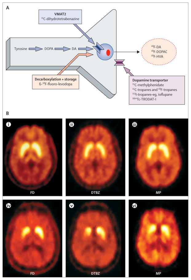
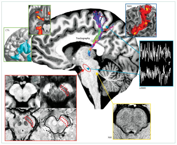
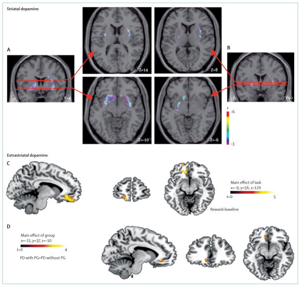
Recent advances in structural and functional imaging have greatly improved our ability to assess normal functions of the basal ganglia, diagnose parkinsonian syndromes, understand the pathophysiology of parkinsonism and other movement disorders, and detect and monitor disease progression. Radionuclide imaging is the best way to detect and monitor dopamine deficiency, and will probably continue to be the best biomarker for assessment of the effects of disease-modifying therapies. However, advances in magnetic resonance enable the separation of patients with Parkinson's disease from healthy controls, and show great promise for differentiation between Parkinson's disease and other akinetic-rigid syndromes. Radionuclide imaging is useful to show the dopaminergic basis for both motor and behavioural complications of Parkinson's disease and its treatment, and alterations in non-dopaminergic systems. Both PET and MRI can be used to study patterns of functional connectivity in the brain, which is disrupted in Parkinson's disease and in association with its complications, and in other basal-ganglia disorders such as dystonia, in which an anatomical substrate is not otherwise apparent. Functional imaging is increasingly used to assess underlying pathological processes such as neuroinflammation and abnormal protein deposition. This imaging is another promising approach to assess the effects of treatments designed to slow disease progression.
Copyright © 2014 Elsevier Ltd. All rights reserved.
Conflict of interest statement
We declare no competing interests.
Figures

References
-
- Kwon DH, Kim JM, Oh SH, et al. Seven-Tesla magnetic resonance images of the substantia nigra in Parkinson disease. Ann Neurol. 2012;71:267–77. - PubMed
-
- Martin WR, Wieler M, Gee M. Midbrain iron content in early Parkinson disease: a potential biomarker of disease status. Neurology. 2008;70:1411–17. - PubMed
-
- Péran P, Cherubini A, Assogna F, et al. Magnetic resonance imaging markers of Parkinson’s disease nigrostriatal signature. Brain. 2010;133:3423–33. - PubMed
-
- Menke RA, Scholz J, Miller KL, et al. MRI characteristics of the substantia nigra in Parkinson’s disease: a combined quantitative T1 and DTI study. Neuroimage. 2009;47:435–41. - PubMed
Publication types
MeSH terms
Substances
Grants and funding
LinkOut - more resources
Full Text Sources
Other Literature Sources
Medical
