. 2014 Jun 23:5:4150.
doi: 10.1038/ncomms5150.

Identification of promiscuous ene-reductase activity by mining structural databases using active site constellations

Affiliations
Free PMC article

Identification of promiscuous ene-reductase activity by mining structural databases using active site constellations

Georg Steinkellner et al. Nat Commun. .
Free PMC article

Abstract

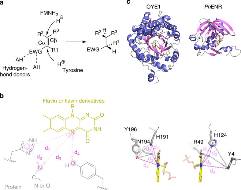
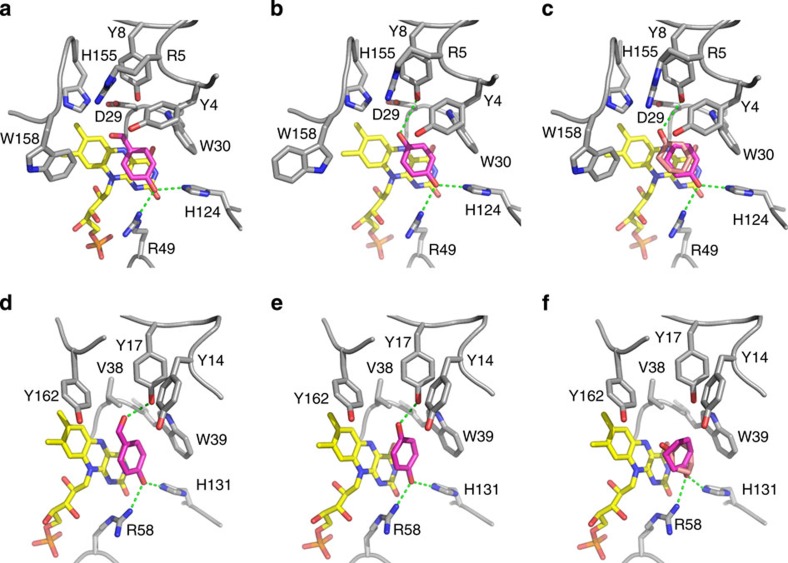
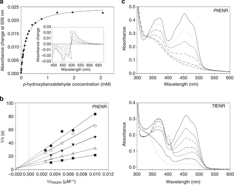
The exploitation of catalytic promiscuity and the application of de novo design have recently opened the access to novel, non-natural enzymatic activities. Here we describe a structural bioinformatic method for predicting catalytic activities of enzymes based on three-dimensional constellations of functional groups in active sites ('catalophores'). As a proof-of-concept we identify two enzymes with predicted promiscuous ene-reductase activity (reduction of activated C-C double bonds) and compare them with known ene-reductases, that is, members of the Old Yellow Enzyme family. Despite completely different amino acid sequences, overall structures and protein folds, high-resolution crystal structures reveal equivalent binding modes of typical Old Yellow Enzyme substrates and ligands. Biochemical and biocatalytic data show that the two enzymes indeed possess ene-reductase activity and reveal an inverted stereopreference compared with Old Yellow Enzymes for some substrates. This method could thus be a tool for the identification of viable starting points for the development and engineering of novel biocatalysts.


Figures

Figure 1
Figure 1. Overview of search template and comparison between OYEs and the identified hits.
(a) Hydrogenation of activated C–C double bonds catalysed by OYEs (EWG, electron withdrawing group; HA, hydrogen bonding donor). (b) Search template with distance descriptors derived from OYE structures. Amino acids of the protein and the flavin cofactor are represented in grey and yellow, respectively. Spheres represent atoms/pseudo-atoms used for defining distance ranges (d1=7.5–7.9 Å, d2=4.6–5.4 Å, d3=6.1–6.8 Å, d4=5.0–8.9 Å). Undefined bond valences are indicated by dotted lines. (c) Comparison of the overall and active site structures of OYE1 (PDB entry: 1OYB) from Saccharomyces pastorianus as a typical member of the OYE family and one protomer of PhENR (PDB entry: 2R6V) identified by the catalophore search. The lower panel shows the positions of the catalytic residues relative to FMN. Distance descriptors d1d4 are shown as magenta dotted lines. Additional grey dotted lines complete a tetrahedron to better show the approximate mirror symmetry.
Figure 2
Figure 2. X-ray crystal structures of ligand complexes of PhENR and TtENR.
Complexes of PhENR with p-hydroxybenzaldehyde (a), 1,4-hydroquinone (b) and 2-cyclohexen-1-one (c). Complexes of TtENR with p-hydroxybenzaldehyde (d), 1,4-hydroquinone (e) and 2-cyclohexen-1-one (f). Amino acid residues are shown in grey, the flavin cofactor in yellow and the bound ligands in magenta. The second, alternate binding mode of 2-cyclohexen-1-one is shown in pink. Hydrogen bonding interactions are depicted as green, dashed lines. The figure was prepared using PyMOL (Schrodinger Inc.).
Figure 3
Figure 3. Biochemical and spectroscopic analysis.
(a) Difference titration of TtENR with p-hydroxybenzaldehyde. The determined Kd value was 25 μM. The insert shows the difference ultraviolet–visible spectra. (b) Double-reciprocal plot of initial rate measurements with PhENR as a function of NADH at 20 (filled circles), 30 (open circles), 50 (filled triangles), 90 (open triangles) and 150 (filled squares) μM of 1,4-benzoquinone. (c) Ultraviolet–visible spectra of anaerobic photoreduction of PhENR (upper panel) and TtENR (lower panel), where the peak at 402 nm indicates the presence of the red radical species.

References

    1. Huisman G. W. & Collier S. J. On the development of new biocatalytic processes for practical pharmaceutical synthesis. Curr. Opin. Chem. Biol. 17, 284–292 (2013). - PubMed
    1. McGrath B. M. & Walsh G. Directory of Therapeutic Enzymes CRC Press (2006).
    1. Turner N. J. Directed evolution drives the next generation of biocatalysts. Nat. Chem. Biol. 5, 567–573 (2009). - PubMed
    1. Furnham N. et al. The Catalytic Site Atlas 2.0: Cataloging catalytic sites and residues identified in enzymes. Nucleic Acids Res. 42, D485–D489 (2014). - PMC - PubMed
    1. Krissinel E. & Henrick K. Secondary-structure matching (SSM), a new tool for fast protein structure alignment in three dimensions. Acta Crystallogr. D 60, 2256–2268 (2004). - PubMed

Publication types