Mapping the development of the basal ganglia in children with attention-deficit/hyperactivity disorder
- PMID: 24954827
- PMCID: PMC10461726
- DOI: 10.1016/j.jaac.2014.05.003
Mapping the development of the basal ganglia in children with attention-deficit/hyperactivity disorder
Abstract
Objective: The basal ganglia are implicated in the pathophysiology of attention-deficit/hyperactivity disorder (ADHD), but little is known of their development in the disorder. Here, we mapped basal ganglia development from childhood into late adolescence using methods that define surface morphology with an exquisite level of spatial resolution.
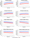
Method: Surface morphology of the basal ganglia was defined from neuroanatomic magnetic resonance images acquired in 270 youth with DSM-IV-defined ADHD and 270 age- and sex-matched typically developing controls; 220 individuals were scanned at least twice. Using linear mixed model regression, we mapped developmental trajectories from age 4 through 19 years at approximately 7,500 surface vertices in the striatum and globus pallidus.
Results: In the ventral striatal surfaces, there was a diagnostic difference in developmental trajectories (t = 5.6, p < .0001). Here, the typically developing group showed surface area expansion with age (estimated rate of increase of 0.54 mm(2) per year, standard error [SE] 0.29 mm(2) per year), whereas the ADHD group showed progressive contraction (decrease of 1.75 mm(2) per year, SE 0.28 mm(2) per year). The ADHD group also showed significant, fixed surface area reductions in dorsal striatal regions, which were detected in childhood at study entry and persisted into adolescence. There was no significant association between history of psychostimulant treatment and developmental trajectories.
Conclusions: Progressive, atypical contraction of the ventral striatal surfaces characterizes ADHD, localizing to regions pivotal in reward processing. This contrasts with fixed, nonprogressive contraction of dorsal striatal surfaces in regions that support executive function and motor planning.
Keywords: attention-deficit/hyperactivity disorder; basal ganglia; development; neuroimaging; ventral striatum.
Published by Elsevier Inc.
Conflict of interest statement
Disclosure: Drs. Shaw, De Rossi, Greenstein, Raznahan, Lerch, and Chakravarty, and Mss. Watson, Wharton, and Sharp report no biomedical financial interests or potential conflicts of interest.
Figures


Comment in
-
Here/In this issue and there/abstract thinking: "80 billion dollars, every year".J Am Acad Child Adolesc Psychiatry. 2014 Jul;53(7):711-2. doi: 10.1016/j.jaac.2014.04.016. J Am Acad Child Adolesc Psychiatry. 2014. PMID: 24954817 No abstract available.
References
-
- Sonuga-Barke EJ. Causal models of attention-deficit/hyperactivity disorder: from common simple deficits to multiple developmental pathways. Biol Psychiatry. 2005;57(11):1231–1238. - PubMed
-
- Sagvolden T, Johansen EB, Aase H, Russell VA. A dynamic developmental theory of attention-deficit/hyperactivity disorder (ADHD) predominantly hyperactive/impulsive and combined subtypes. Behavioral and Brain Sciences. 2005;28(3):397–418. - PubMed
-
- Willcutt EG, Doyle AE, Nigg JT, Faraone SV, Pennington BF. Validity of the executive function theory of attention-deficit/hyperactivity disorder: a meta-analytic review. Biol Psychiatry. Jun 1 2005;57(11):1336–1346. - PubMed
-
- Barkley RA. Behavioral inhibition, sustained attention, and executive functions: constructing a unifying theory of ADHD. Psychological Bulletin. Jan 1997;121(1):65–94. - PubMed
Publication types
MeSH terms
Grants and funding
LinkOut - more resources
Full Text Sources
Other Literature Sources
Medical

