

Interleukin-6 drives melanoma cell motility through p38α-MAPK-dependent up-regulation of WNT5A expression
- PMID: 24954857
- PMCID: PMC5528610
- DOI: 10.1016/j.molonc.2014.05.008
Interleukin-6 drives melanoma cell motility through p38α-MAPK-dependent up-regulation of WNT5A expression
Abstract
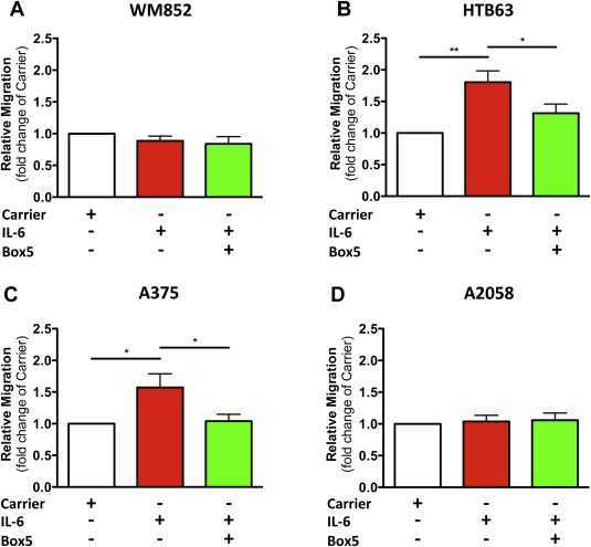
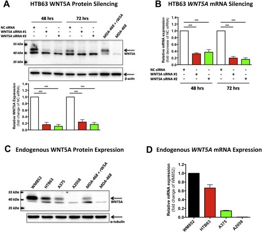
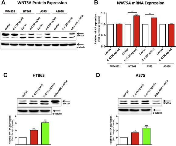
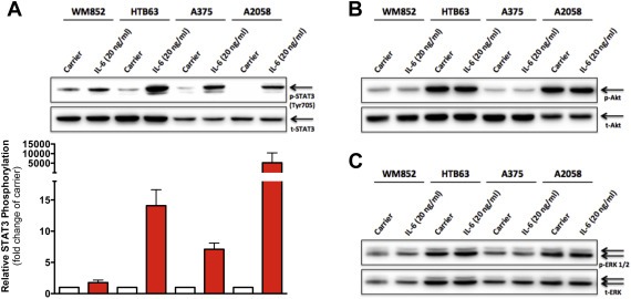
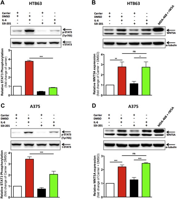
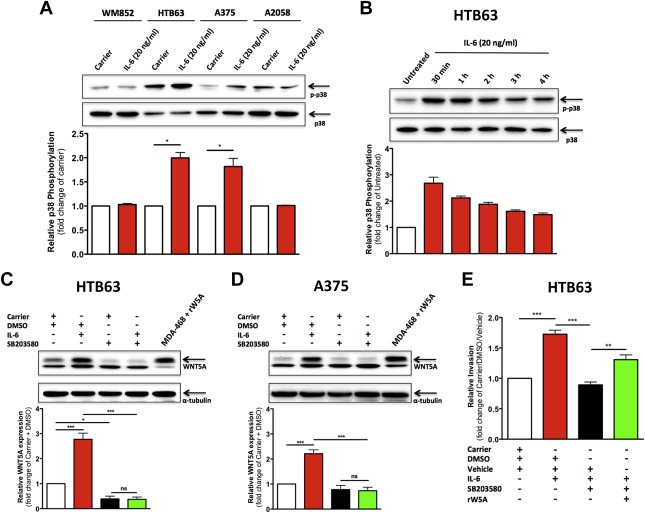
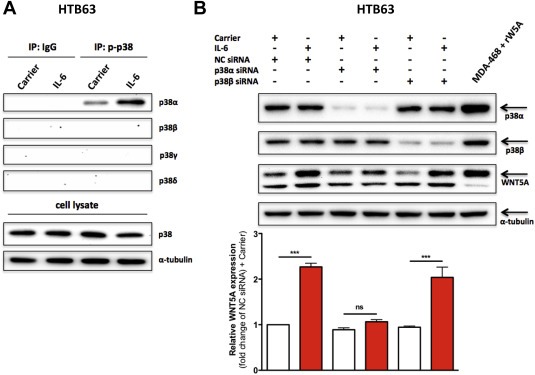
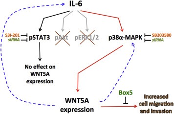
Extensive research has demonstrated a tumor-promoting role of increased WNT5A expression in malignant melanoma. However, very little light has been shed upon how WNT5A expression is up-regulated in melanoma. A potential regulator of WNT5A expression is the pro-inflammatory cytokine Interleukin (IL)-6, which shares the ability of WNT5A to increase melanoma cell invasion. Here, we investigate whether IL-6 can promote melanoma cell motility through an increased expression of WNT5A. We clearly demonstrate that the WNT5A-antagonistic peptide Box5 could inhibit IL-6-induced melanoma cell migration and invasion. Furthermore, IL-6 stimulation of the human melanoma cell lines HTB63 and A375 increased the expression of WNT5A in a dose-dependent manner. To identify the signaling mechanism responsible for this up-regulation, we explored the involvement of the three main signals induced by IL-6; STAT3, Akt and ERK 1/2. Of these, only STAT3 was activated by IL-6 in the melanoma cell lines tested. However, the STAT3 inhibitor S3I-201 failed to inhibit IL-6-induced WNT5A up-regulation in HTB63 and A375 cells. Nor did STAT3 siRNA silencing affect the expression of WNT5A. In search of an alternative signaling mechanism, we detected IL-6-induced activation of p38-MAPK in HTB63 and A375 cells. The p38-MAPK inhibitor SB203580 abolished the IL-6-induced WNT5A up-regulation and blocked IL-6-induced melanoma cell invasion. The latter effect could be rescued by the addition of recombinant WNT5A. Notably, immunoprecipitation analysis revealed that only the p38α-MAPK isoform was activated by IL-6, and subsequent siRNA silencing of p38α-MAPK abolished the IL-6-induced up-regulation of WNT5A. Taken together, we demonstrate a novel link between the two melanoma pro-metastatic agents IL-6 and WNT5A explaining how IL-6 can increase melanoma cell invasion and thus promote the metastatic process. This finding provides a basis for future therapeutic intervention of melanoma progression.
Keywords: Cell motility; Interleukin-6; Melanoma; STAT3; WNT5A; p38 MAPK.
Copyright © 2014 Federation of European Biochemical Societies. Published by Elsevier B.V. All rights reserved.
Figures
Similar articles
-
A t-butyloxycarbonyl-modified Wnt5a-derived hexapeptide functions as a potent antagonist of Wnt5a-dependent melanoma cell invasion.Proc Natl Acad Sci U S A. 2009 Nov 17;106(46):19473-8. doi: 10.1073/pnas.0909409106. Epub 2009 Nov 9. Proc Natl Acad Sci U S A. 2009. PMID: 19901340 Free PMC article.
-
WNT5A induces release of exosomes containing pro-angiogenic and immunosuppressive factors from malignant melanoma cells.Mol Cancer. 2014 Apr 26;13:88. doi: 10.1186/1476-4598-13-88. Mol Cancer. 2014. PMID: 24766647 Free PMC article.
-
Migration and invasion of oral squamous carcinoma cells is promoted by WNT5A, a regulator of cancer progression.J Oral Pathol Med. 2015 Nov;44(10):776-84. doi: 10.1111/jop.12292. Epub 2014 Dec 2. J Oral Pathol Med. 2015. PMID: 25459554
-
Phenylmethimazole decreases Toll-like receptor 3 and noncanonical Wnt5a expression in pancreatic cancer and melanoma together with tumor cell growth and migration.Clin Cancer Res. 2009 Jun 15;15(12):4114-22. doi: 10.1158/1078-0432.CCR-09-0005. Epub 2009 May 26. Clin Cancer Res. 2009. PMID: 19470740 Free PMC article.
-
The Role of Melanoma Cell-Stroma Interaction in Cell Motility, Invasion, and Metastasis.Front Med (Lausanne). 2018 Nov 6;5:307. doi: 10.3389/fmed.2018.00307. eCollection 2018. Front Med (Lausanne). 2018. PMID: 30460237 Free PMC article. Review.
Cited by
-
WNT5A signaling impairs breast cancer cell migration and invasion via mechanisms independent of the epithelial-mesenchymal transition.J Exp Clin Cancer Res. 2016 Sep 13;35(1):144. doi: 10.1186/s13046-016-0421-0. J Exp Clin Cancer Res. 2016. PMID: 27623766 Free PMC article.
-
Correlations between peripheral blood biomarkers and clinical outcomes in advanced non-small cell lung cancer patients who received immunotherapy-based treatments.Transl Lung Cancer Res. 2021 Dec;10(12):4477-4493. doi: 10.21037/tlcr-21-710. Transl Lung Cancer Res. 2021. PMID: 35070755 Free PMC article.
-
WNT-5A: signaling and functions in health and disease.Cell Mol Life Sci. 2016 Feb;73(3):567-87. doi: 10.1007/s00018-015-2076-y. Epub 2015 Oct 29. Cell Mol Life Sci. 2016. PMID: 26514730 Free PMC article. Review.
-
Trained immunity alleviates the progression of melanoma during sepsis-associated immunoparalysis.Cell Oncol (Dordr). 2025 Aug;48(4):1047-1065. doi: 10.1007/s13402-025-01063-8. Epub 2025 Apr 9. Cell Oncol (Dordr). 2025. PMID: 40205307 Free PMC article.
-
Frizzled Receptors in Tumors, Focusing on Signaling, Roles, Modulation Mechanisms, and Targeted Therapies.Oncol Res. 2021 Mar 16;28(6):661-674. doi: 10.3727/096504020X16014648664459. Epub 2020 Sep 30. Oncol Res. 2021. PMID: 32998794 Free PMC article. Review.
References
-
- Bergenfelz, C. , Janols, H. , Wullt, M. , Jirstrom, K. , Bredberg, A. , Leandersson, K. , 2013. Wnt5a inhibits human monocyte-derived myeloid dendritic cell generation. Scand. J. Immunol.. 78, 194–204. - PubMed
-
- Bittner, M. , Meltzer, P. , Chen, Y. , Jiang, Y. , Seftor, E. , Hendrix, M. , Radmacher, M. , Simon, R. , Yakhini, Z. , Ben-Dor, A. , Sampas, N. , Dougherty, E. , Wang, E. , Marincola, F. , Gooden, C. , Lueders, J. , Glatfelter, A. , Pollock, P. , Carpten, J. , Gillanders, E. , Leja, D. , Dietrich, K. , Beaudry, C. , Berens, M. , Alberts, D. , Sondak, V. , 2000. Molecular classification of cutaneous malignant melanoma by gene expression profiling. Nature. 406, 536–540. - PubMed
-
- Da Forno, P.D. , Pringle, J.H. , Hutchinson, P. , Osborn, J. , Huang, Q. , Potter, L. , Hancox, R.A. , Fletcher, A. , Saldanha, G.S. , 2008. WNT5A expression increases during melanoma progression and correlates with outcome. Clin. Cancer Res. : An Off. J. Am. Assoc. Cancer Res.. 14, 5825–5832. - PubMed
Publication types
MeSH terms
Substances
LinkOut - more resources
Full Text Sources
Other Literature Sources
Medical
Molecular Biology Databases
Miscellaneous
