. 2014 Jul 3;55(1):111-22.
doi: 10.1016/j.molcel.2014.05.020. Epub 2014 Jun 19.

Co-opting the Fanconi anemia genomic stability pathway enables herpesvirus DNA synthesis and productive growth

Affiliations

Co-opting the Fanconi anemia genomic stability pathway enables herpesvirus DNA synthesis and productive growth

Heidi Karttunen et al. Mol Cell. .

Abstract

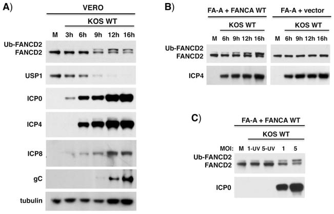
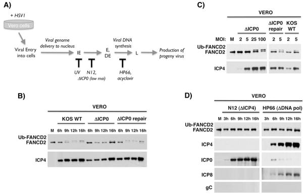
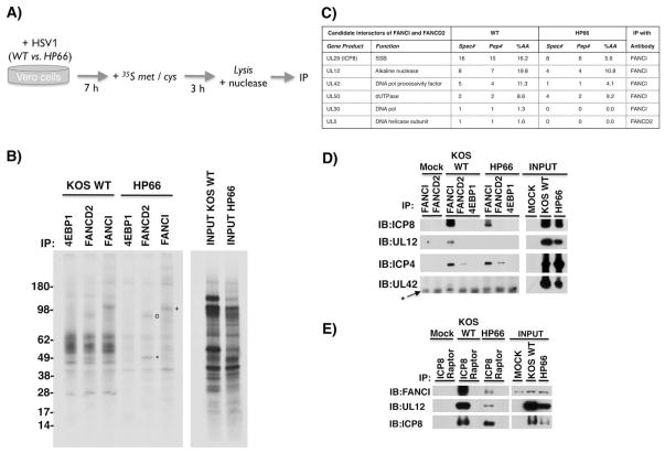
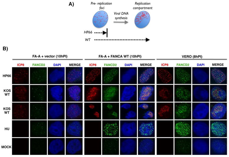
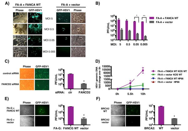
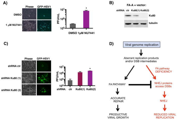
DNA damage associated with viral DNA synthesis can result in double-strand breaks that threaten genome integrity and must be repaired. Here, we establish that the cellular Fanconi anemia (FA) genomic stability pathway is exploited by herpes simplex virus 1 (HSV-1) to promote viral DNA synthesis and enable its productive growth. Potent FA pathway activation in HSV-1-infected cells resulted in monoubiquitination of FA effector proteins FANCI and FANCD2 (FANCI-D2) and required the viral DNA polymerase. FANCD2 relocalized to viral replication compartments, and FANCI-D2 interacted with a multisubunit complex containing the virus-encoded single-stranded DNA-binding protein ICP8. Significantly, whereas HSV-1 productive growth was impaired in monoubiquitination-defective FA cells, this restriction was partially surmounted by antagonizing the DNA-dependent protein kinase (DNA-PK), a critical enzyme required for nonhomologous end-joining (NHEJ). This identifies the FA-pathway as a cellular factor required for herpesvirus productive growth and suggests that FA-mediated suppression of NHEJ is a fundamental step in the viral life cycle.


Figures

Figure 1
Figure 1. Activation of the FA pathway in HSV1-infected cells
A) Vero cells were mock-infected (M) or infected with WT HSV1 KOS (MOI=5). Total protein was harvested at different times post-infection (hPI), fractionated by SDS-PAGE and analyzed by immunoblotting using the indicated antibodies. Ub-FANCD2 denotes the slower- migrating, monoubiquitinated FANCD2. B) Patient-derived FA-A cells (FANCA-deficient) stably transduced with either empty expression vector only (FA-A vector) or functionally complemented with a WT FANCA cDNA (FA-A + FANCA WT) and treated as in A. C) FA-A cells complemented with FANCA WT were mock-infected (M) or infected with WT or UV-inactivated WT HSV1 KOS (MOI=1 or 5). Total protein harvested at 19 hpi was analyzed as in A.
Figure 2
Figure 2. FA pathway activation in HSV1-infected cells requires viral gene expression
A) Temporal progression of the HSV1 productive replication cycle. Upon HSV1-infection of permissive Vero cells, the virus enters cells and the dsDNA viral genome is delivered to the nucleus. The viral gene expression program commences with transcription of immediate early (IE) genes, which in turn activate transcription of early (E) and delayed-early (DE) genes. This is followed by viral DNA synthesis, which activates transcription of late (L) viral genes, and culminates in infectious progeny virus production. The point at which different mutant viruses (ΔICP0 at low MOI, N12, HP66) or treatments (UV irradiated virus, acyclovir) arrest the viral lifecycle is shown below. B) Vero cells were mock-infected (M) or infected with WT KOS, ΔICP0 or ΔICP0-repair viruses (MOI = 5). Total protein was harvested at various times (hPI), fractionated by SDS-PAGE and analyzed by immunoblotting using the indicated antibodies. C) As in A except cells were mock-infected or infected at the indicated MOI (2, 5, 25,100). After 9 hpi, total protein was isolated and analyzed as in B. D) Vero cells were infected (MOI=5) with a virus deficient for ICP4 (N12) or the viral DNA Pol (HP66). Total protein isolated at the indicated time was analyzed as in B. In addition to FANCD2, accumulation of representative viral IE (ICP4, ICP0), early (ICP8) and late (gC) proteins were monitored. ICP0 is overexpressed in the absence of ICP4 (DeLuca et al., 1985), and preventing DNA synthesis limits ICP0 expression (Weinheimer & McKnight, 1987).
Figure 3
Figure 3. Association of FANCD2-FANCI with specific virus-encoded proteins in HSV1-infected cells
A) Experimental plan to identify HSV1 polypeptides associated with cellular FA proteins. Vero cells were infected (MOI=10) with either WT HSV1 or a mutant virus that arrests its growth cycle prior to initiating DNA synthesis (HP66). After 7h, cultures were metabolically labeled with 35S met / cys for 3h. Under these conditions, host protein synthesis is impaired and the vast majority of newly synthesized, radiolabeled polypeptides are HSV1-encoded. A cell-free lysate prepared using non-ionic detergent was nuclease-treated to degrade single and ds nucleic acid, and subsequently IP’ed using anti-FANCD2, anti-FANCI, or a control affinity-purified rabbit antisera that recognizes an unrelated protein (the translational repressor 4E-BP1). B) Immune complexes isolated from infected, metabolically-labeled lysates described and treated as in A were fractionated by SDS-PAGE and analyzed by autoradiography. A sample of input lysate is shown (right panel). Radiolabeled proteins with molecular weights of 130–150 kDA (+), 80 kDA (o), and 50 kDA (*) are indicated on the autoradiogram. Molecular weight standards with their indicated relative molecular weight (kDa) appear to the left. C) Anti-FANCI and anti-FANCD2 immune complexes isolated from HSV1-infected (WT vs. HP66) cells were analyzed by tandem MS. HSV1-encoded DNA replication and repair proteins (gene products) associated with FANCI-D2, the number of spectral counts (Spec#), the number of peptides identified (Pep#), % amino acid coverage (%AA), and the antibody used for IP are shown. D) Non-ionic detergent lysates prepared from Vero cells mock-infected (M) or infected as in A were nuclease-treated and IP’ed using the specified antisera. Immune complexes were fractionated by SDS-PAGE and analyzed by immunoblotting (IB) using the indicated antisera. A sample of input lysate is shown (right panel). * indicates IgG heavy chain. E) As in D except samples were IP’ed using anti-ICP8 or an isotype matched control antisera (anti-raptor).
Figure 4
Figure 4. Redistribution of FANCD2 in response to HSV1-infection and accumulation proximal to nuclear viral replication compartments (RCs)
A) Maturation of HSV1 pre-replication foci into nuclear RCs is dependent upon viral DNA synthesis. The HSV1-encoded SSB ICP8 is one of several viral replication proteins that accumulate at discrete foci (depicted as red dots) distributed within host nuclei (blue). Viral DNA synthesis triggers rearrangement of replication foci into an organized RC within nuclei. Since the HP66 mutant lacks a functional viral DNA pol and cannot replicate viral DNA, ICP8 only accumulates in pre-replication foci and the viral lifecycle arrests at this point. Pre-replication foci mature into RCs in cells infected with WT virus. B) Vero cells or FA-A cells stably transduced with either empty expression vector only (FA-A + vector) or a WT FANCA cDNA (FA-A + FANCA WT) were mock-infected or infected (MOI=5) with HP66 or WT KOS (10 h for FA-A cells; 8 h for Vero cells) and subsequently processed for indirect immunofluorescence. As a positive control to detect FANCD2 nuclear foci after DNA damage, cells were treated with 2 mM hydroxyurea (HU) for 8 or 10 h. Cells were co-stained to detect either ICP8 (red), FANCD2 (green), or DAPI (blue) and a representative image for each condition is shown.
Figure 5
Figure 5. The cellular FA pathway is required for productive HSV1 replication
A) FA-A cells stably transduced with either empty expression vector only (FA-A + vector, right panel) or complemented with a WT FANCA cDNA (FA-A + FANCA WT, left panel) were infected at the indicated MOI with the WT EGFP-HSV1 reporter strain (GFP-HSV1). This strain contains EGFP-fused to the virus Us11 late gene and is indistinguishable from WT virus in its replicative capacity (Benboudjema et al., 2003). After 3 d, live cells were evaluated by phase contrast and epifluorescence microscopy. Images of representative fields are shown. B) Infectious virus produced in the experiment shown in panel A was quantified by plaque assay in Vero cells. * p=0.0098; ** p=0.00001. C) Primary human fibroblasts transfected with non-silencing, control siRNA or FANCD2 siRNA (Fig. S5) were infected with GFP-HSV1 (MOI = 10−3) and evaluated as in A after 2 d post-infection (Left panel). Infectious virus produced was quantified as in B (Right panel). * p=0.0282. D) FA-A + vector or FA-A + FANCA WT cells were infected (MOI=5) with HP66 or WT KOS, and total genomic DNA was isolated at the indicated times. Relative viral genomic DNA levels were determined by qPCR using primers complementary to the HSV1 ICP27 gene. Input DNA was normalized by amplifying the RPL19 gene. E) FA-G cells stably transduced with either empty expression vector only (FA-G + vector) or functionally complemented with a WT FANCG cDNA (FA-G + FANCG WT) were infected (MOI= 0.5 × 10⁻³) with the WT EGFP-HSV1 reporter strain (GFP-HSV1). After 2 d, live cells were evaluated by microscopy as in A (Left panel). Infectious virus produced was quantified as in B (Right panel). * p=0.001. F) BRCA2-deficient cells stably transfected with empty expression vector (BRCA2 vector) or functionally reconstituted with chromosome 13, which contains a WT BRCA2 gene (BRCA2 WT), were infected (MOI= 5 × 10⁻³) and analyzed as in E. * p=0.009
Figure 6
Figure 6. Inhibiting DNA-PKcs partially restores HSV-1 replication in FA-deficient cells
A) FA-A cells stably transduced with empty expression vector only (FA-A + vector) were infected with EGFP-HSV-1 (MOI = 0.05) in the presence of the DNA-PKcs inhibitor NU7441 (1μM) or vehicle control (DMSO). After 3 d, live cells were evaluated by phase contrast and epifluorescence microscopy. Images of representative fields are shown for three separate experiments (left panel). Infectious virus produced was quantified by plaque assay in Vero cells (right panel). * p=0.0101. B) FA-A cells as in A were transduced with a lentivirus expressing control, non-silencing shRNA (ctr), or one of two shRNAs targeting Ku80 (Ku80(1), Ku80(2)). After 4 d, total protein was harvested and the abundance of Ku80 vs tubulin (loading control) evaluated by immunoblotting. C). As in B except cells transduced with the indicated shRNA-expressing lentivirus were infected with GFP-HSV1 (MOI= 5 × 10⁻²). After 6 d, live cells were evaluated by microscopy (left panel) and infectious virus quantified (right panel) as in A. * p= 0.0003. D) Model depicting the proposed role of the FA pathway in counteracting aberrant NHEJ activity during HSV1 genome replication. Viral infection co-opts the cellular FA pathway by stimulating redistribution and monoubiquitination of FA effector proteins, FANCI and FANCD2, near viral replication sites. The precise role of monoubiquitinated FANCI-D2 at these sites remains unclear but may involve counteracting illegitimate DNA transactions by NHEJ during viral replication. Thus, the cellular FA pathway is subverted by HSV1 to enable efficient viral genome replication and promote productive viral growth.

Similar articles

Cited by

References

    1. Adamo A, Collis SJ, Adelman CA, Silva N, Horejsi Z, Ward JD, Martinez-Perez E, Boulton SJ, La Volpe A. Preventing non-homologous end joining suppresses DNA repair defects of Fanconi anemia. Mol Cell. 2010;39:25–35. - PubMed
    1. Balasubramanian N, Bai P, Buchek G, Korza G, Weller SK. Physical interaction between the herpes simplex virus type 1 exonuclease, UL12, and the DNA double-strand break-sensing MRN complex. J Virol. 2010;84:12504–12514. - PMC - PubMed
    1. Benboudjema L, Mulvey M, Gao Y, Pimplikar SW, Mohr I. Association of the herpes simplex virus type 1 Us11 gene product with the cellular kinesin light-chain-related protein PAT1 results in the redistribution of both polypeptides. J Virol. 2003;77:9192–9203. - PMC - PubMed
    1. Boichuk S, Hu L, Hein J, Gjoerup OV. Multiple DNA damage signaling and repair pathways deregulated by simian virus 40 large T antigen. J Virol. 2010;84:007–8020. - PMC - PubMed
    1. Chapman JR, Taylor MR, Boulton SJ. Playing the end game: DNA double-strand break repair pathway choice. Mol Cell. 2012;47:497–510. - PubMed

Publication types

MeSH terms

Substances