

False-positive reduction in mammography using multiscale spatial Weber law descriptor and support vector machines
- PMID: 24954976
- PMCID: PMC4055841
- DOI: 10.1007/s00521-013-1450-7
False-positive reduction in mammography using multiscale spatial Weber law descriptor and support vector machines
Abstract
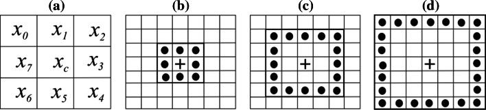
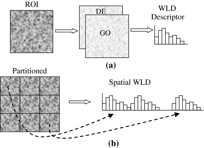
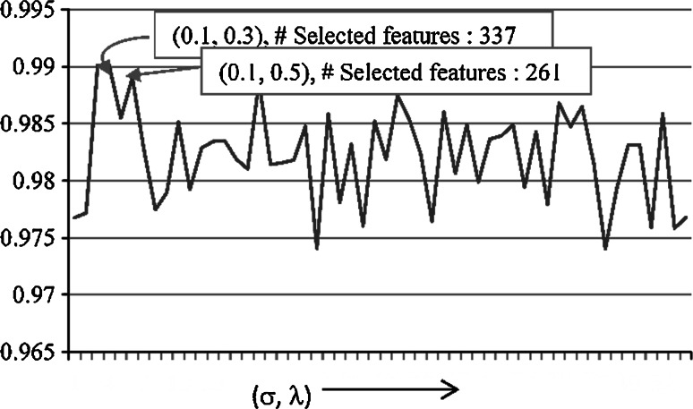
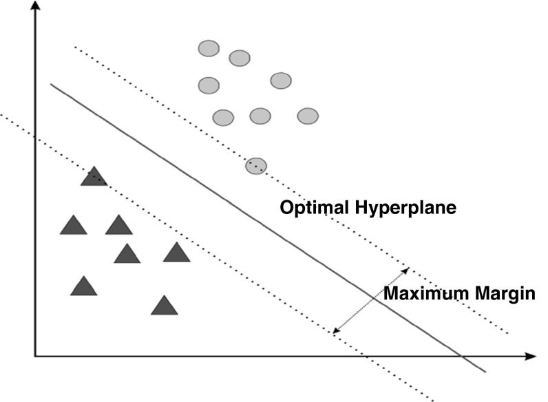
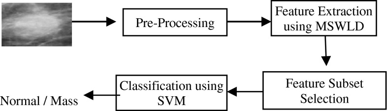
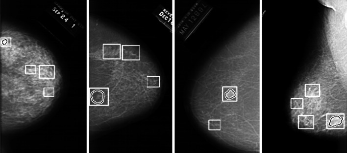
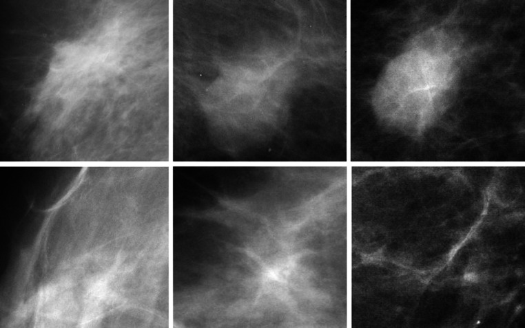
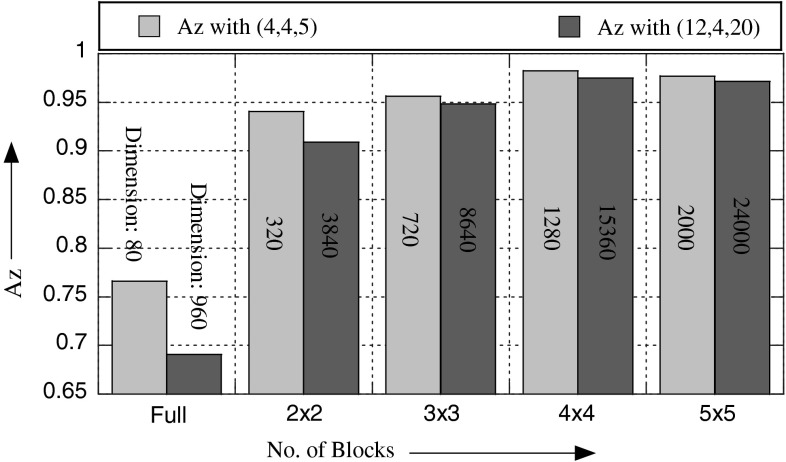
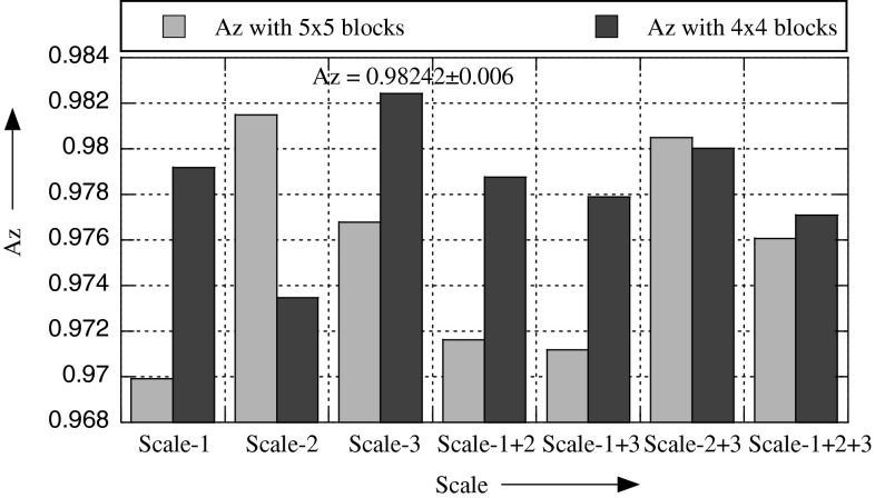
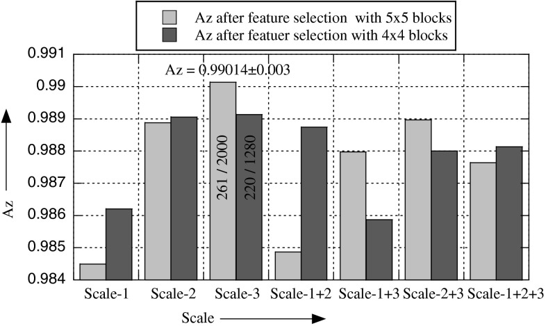
In a CAD system for the detection of masses, segmentation of mammograms yields regions of interest (ROIs), which are not only true masses but also suspicious normal tissues that result in false positives. We introduce a new method for false-positive reduction in this paper. The key idea of our approach is to exploit the textural properties of mammograms and for texture description, to use Weber law descriptor (WLD), which outperforms state-of-the-art best texture descriptors. The basic WLD is a holistic descriptor by its construction because it integrates the local information content into a single histogram, which does not take into account the spatial locality of micropatterns. We extend it into a multiscale spatial WLD (MSWLD) that better characterizes the texture micro structures of masses by incorporating the spatial locality and scale of microstructures. The dimension of the feature space generated by MSWLD becomes high; it is reduced by selecting features based on their significance. Finally, support vector machines are employed to classify ROIs as true masses or normal parenchyma. The proposed approach is evaluated using 1024 ROIs taken from digital database for screening mammography and an accuracy of Az = 0.99 ± 0.003 (area under receiver operating characteristic curve) is obtained. A comparison reveals that the proposed method has significant improvement over the state-of-the-art best methods for false-positive reduction problem.
Keywords: False-positive reduction; Mammograms; Mass detection; Support vector machines; WLD.
Figures
References
-
- Mu T, Nandi AK (2005) Detection of breast cancer using v-SVM and RBF networks with self-organization selection of centers. In: Third IEEE international seminar on medical applications of signal processing
-
- American Cancer Society (2003–2004) Breast cancer: facts and figures. ACS
-
- Esteve J, Kricker A, Ferlay J, Parkin D (1993) Facts and figures of cancer in the European Community. In: Technical report on international agency for research on cancer
-
- Pal NR, Bhowmick B, Patel SK, Pal S, Das J. A multi-stage neural network aided system for detection of micro-calcifications in digitized mammograms. Neurocomputing. 2008;71:2625–2634. doi: 10.1016/j.neucom.2007.06.015. - DOI
LinkOut - more resources
Full Text Sources
Other Literature Sources
Miscellaneous