. 2014 Jun 10;4(8):845-57.
doi: 10.7150/thno.8343. eCollection 2014.

MRI of breast tumor initiating cells using the extra domain-B of fibronectin targeting nanoparticles

Affiliations

MRI of breast tumor initiating cells using the extra domain-B of fibronectin targeting nanoparticles

Yujin Sun et al. Theranostics. .

Abstract

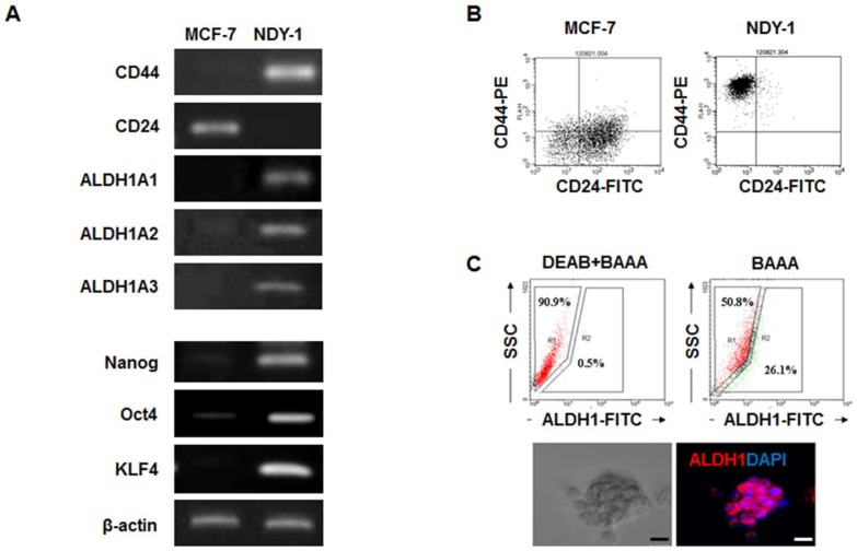
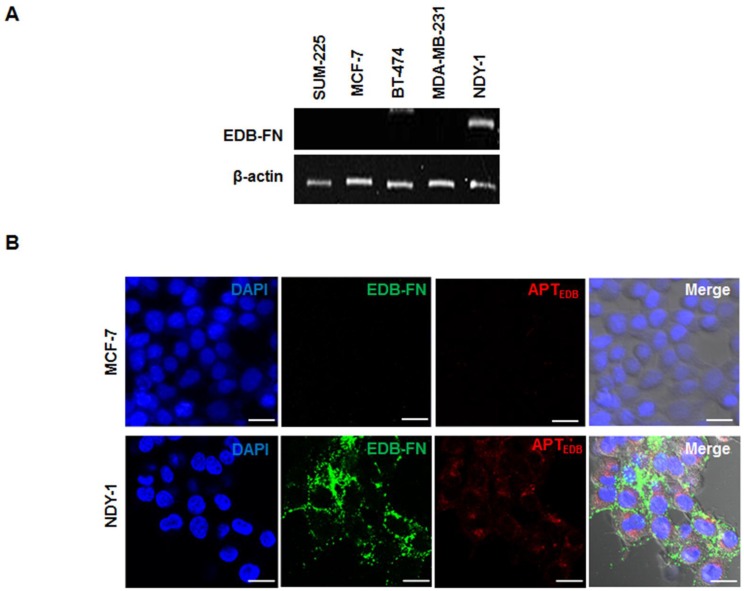
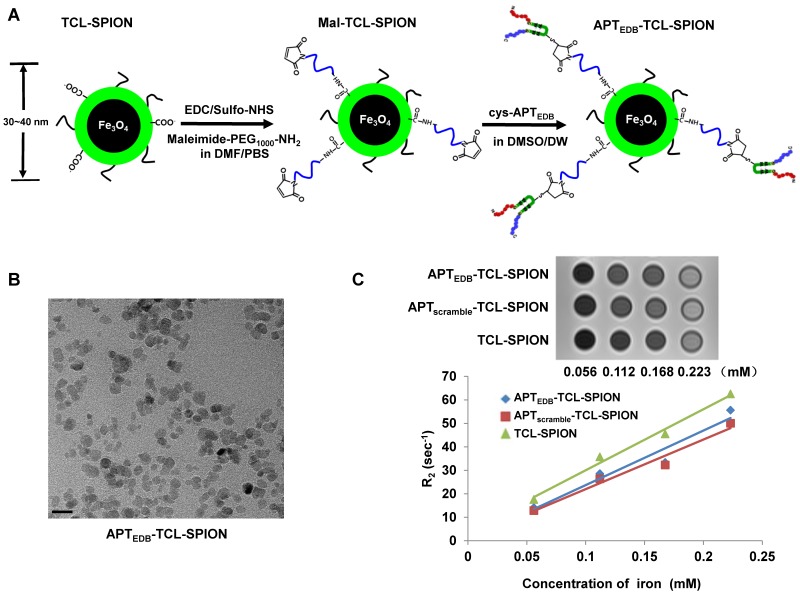
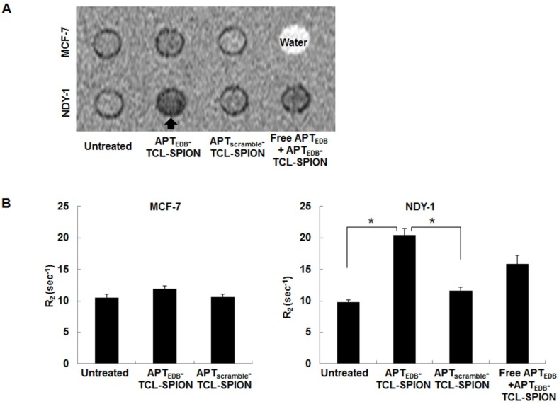
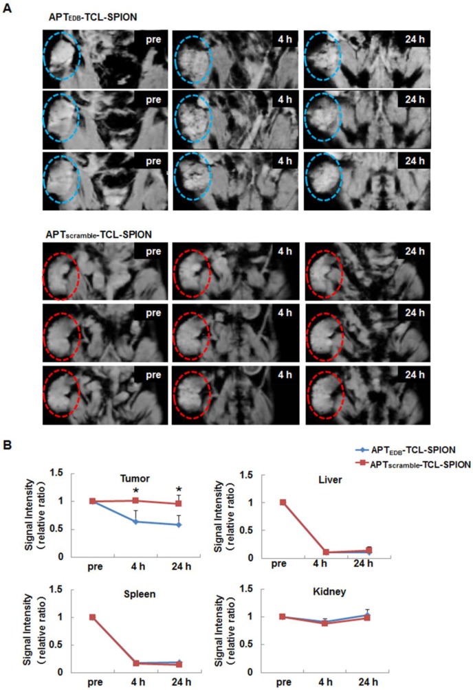
The identification of breast tumor initiating cells (BTICs) is important for the diagnosis and therapy of breast cancers. This study was undertaken to evaluate whether the extra domain-B of fibronectin (EDB-FN) could be used as a new biomarker for BTICs and whether EDB-FN targeting superparamagnetic iron oxide nanoparticles (SPIONs) could be used as a magnetic resonance imaging (MRI) contrast agent for BTIC imaging in vitro and in vivo. BTICs (NDY-1) exhibited high EDB-FN expression, whereas non-BTICs (MCF-7, BT-474, SUM-225, MDA-MB-231) did not exhibit EDB-FN expression. Furthermore, Cy3.3-labeled EDB-FN specific peptides (APTEDB) showed preferential binding to the targeted NDY-1 cells. To construct an EDB-FN targeted imaging probe, APTEDB was covalently attached to a thermally cross-linked SPION (TCL-SPION) to yield APTEDB-TCL-SPION. In the in vitro MRI of cell phantoms, selective binding of APTEDB-TCL-SPION to NDY-1 cells was evident, but little binding was observed in MCF-7 cells. After the intravenous injection of APTEDB-TCL-SPION into the NDY-1 mouse tumor xenograft model, a significant decrease in the signal within the tumor was observed in the T2*-weighted images; however, there was only a marginal change in the signal of non-targeting SPIONs such as APTscramble-TCL-SPION or TCL-SPION. Taken together, we report for the first time that EDB-FN was abundantly expressed in BTICs and may therefore be useful as a new biomarker for identifying BTICs. Our study also suggests that APTEDB-TCL-SPION could be used as an MRI contrast agent for BTIC imaging.

Keywords: Aptides; Breast tumor initiating cells; Extra domain-B of fibronectin; Magnetic resonance imaging.; Superparamagnetic iron oxide nanoparticles.


Conflict of interest statement

Competing Interests: The authors have declared that no competing interest exists.

Figures

Figure 1
Figure 1
Analysis of the genes expressed in breast tumor initiating cells (BTICs). (A) RT-PCR analysis of the self-renewal genes and surface marker genes in BTICs (NDY-1) and breast cancer cells (MCF-7). Specific markers of BTICs and self-renewal genes were highly expressed in NDY-1 cells but not in MCF-7 cells. (B) Flow cytometry analysis of the surface markers CD44 and CD24. NDY-1 exhibited a CD44+/CD24- BTIC phenotype. (C) ALDEFLUOR assay for ALDH1 activity and Immunostaining analysis for ALDH1 expression. Approximately 26% of the NDY-1 cells exhibited brightly fluorescent ALDH1 staining, indicating the presence of a putative BTIC marker. Immunostaining showed NDY-1 spheroids strongly expressed the ALDH1 protein. Scale bar: 20 μm
Figure 2
Figure 2
Analysis of the expression of extra domain-B of fibronectin (EDB-FN) and its selective targeting with an EDB-FN aptide (APTEDB). (A) RT-PCR analysis of the EDB-FN mRNA in diverse breast cancer cell lines. EDB-FN was expressed in NDY-1 cells but not in SUM-225, MCF-7, BT-474 or MDA-MB-231 cells. (B) EDB-FN targeting images of the Cy3.3-labeled APTEDB (Cy3.3-APTEDB, red fluorescence) and immunostained EDB-FN (green fluorescence) images in breast cancer cells. Cy3.3-APTEDB specific signals were observed in EDB-FN overexpressing NDY-1 cells but not in MCF-7 cells. Scale bar: 20 μm
Figure 3
Figure 3
Synthetic procedures and characterization of APTEDB-TCL-SPION. (A) Schematic illustration of the preparation of APTEDB-TCL-SPIONs. (B) A TEM image of APTEDB-TCL-SPION. Scale bar: 20 nm (C) T2-weighted images and plots of the relaxation rates (R2 =1/T2) of APTEDB-TCL-SPION, APTscramble-TCL-SPION, and TCL-SPION at various iron concentrations. The transverse relaxivities (r2) of APTEDB-TCL-SPION, APTscramble-TCL-SPION and TCL-SPION were 232.1 ± 1.4 mM-1 sec-1, 211.1± 2.1 mM-1 sec-1, and 259.9 ± 3.2 mM-1 sec-1, respectively.
Figure 4
Figure 4
In vitro MRI analysis of cell phantoms. (A) Representative T2-weighted images of cell phantoms. Cells were treated with APTEDB-TCL-SPION and APTscramble-TCL-SPION (11.2 µg Fe/ml) for 12 h at 37°C. Blocking of specific binding of APTEDB-TCL-SPION was achieved by pretreatment with EDB-FN aptides (APTEDB, 0.1 mg/ml) for 1 h. Hypointense signals (arrow) were clearly detected in NDY-1 cells treated with APTEDB-TCL-SPIONs. (B) R2 values measured from the T2-weighted images obtained from cell phantoms. The R2 values significantly increased in the APTEDB-TCL-SPION-treated NDY-1 cells compared with the APTscramble-TCL-SPION-treated or untreated NDY-1 cells. Significantly different R2 values were not observed in APTEDB-TCL-SPION- and APTscramble-TCL-SPION- treated and untreated MCF-7 cells. Pre-incubation with free APTEDB inhibited the increase in R2 values observed for APTEDB-TCL-SPION-treated NDY-1 cells. All values are presented as the mean ± standard deviation of at least three independent experiments. Asterisks (*) indicate that the p value was statistically significant (<0.05).
Figure 5
Figure 5
In vivo EDB-FN targeted tumor MRI. (A) T2*-weighted multi-slice images of the NDY-1 tumor in the mouse obtained prior to injection and at 4 h and 24 h after the injection of APTEDB-TCL-SPION or APTscramble-TCL-SPION (20 mg Fe/kg). The dotted line with circles indicates the engrafted tumor region. Multifocal hypointense spots were observed in tumors obtained from mice injected with APTEDB-TCL-SPION. (B) The signal intensity changes of the tumor areas, liver, spleen and kidney obtained from T2*-weighted images. An apparent signal intensity decrease within the tumor was detected only after APTEDB-TCL-SPION injection. All values are presented as the mean ± standard deviation of at least three independent experiments. Asterisks (*) indicate that the p value was statistically significant (<0.05).
Figure 6
Figure 6
Histological analysis of the tumors. (A) Hematoxylin and eosin (H&E) staining, Prussian blue staining and EDB-FN immunostaining. Prussian blue staining showed that a larger number of accumulated SPIONs were detected as blue dots in the tumors obtained from mice injected with APTEDB-TCL-SPION compared with APTscramble-TCL-SPION. Immunostaining of EDB-FN in NDY-1 tumor microsections was performed using the BC-1 antibody. EDB-FN proteins (dark brown) were abundantly detected in the NDY-1 tumors injected with APTEDB-TCL-SPION or APTscramble-TCL-SPION. (B) Immunostaining of CD31. Immunostaining of the platelet endothelial cell adhesion molecule (CD31) showing that vessels (dark brown) were observed in NDY-1 tumors obtained from mice injected with APTEDB-TCL-SPION. (C) Co-staining of an anti-EDB-FN antibody and Prussian blue. The accumulation of APTEDB-TCL-SPION (blue dots) was observed in EDB-FN positive areas of the tumor vasculature and interstitium. Scale bar: 100 μm

Similar articles

Cited by

References

    1. Al-Hajj M, Wicha MS, Benito-Hernandez A. et al. Prospective identification of tumorigenic breast cancer cells. Proc Natl Acad Sci U S A. 2003;100:3983–8. - PMC - PubMed
    1. Dean M, Fojo T, Bates S. Tumour stem cells and drug resistance. Nat Rev Cancer. 2005;5:275–84. - PubMed
    1. Liu H, Patel MR, Prescher JA. et al. Cancer stem cells from human breast tumors are involved in spontaneous metastases in orthotopic mouse models. Proc Natl Aca Sci U S A. 2010;107:18115–20. - PMC - PubMed
    1. Ginestier C, Hur MH, Charafe-Jauffret E. et al. ALDH1 is a marker of normal and malignant human mammary stem cells and a predictor of poor clinical outcome. Cell Stem Cell. 2007;1:555–67. - PMC - PubMed
    1. Potts JR, Campbell ID. Fibronectin structure and assembly. Curr Opin Cell Biol. 1994;6:648–55. - PubMed

Publication types

MeSH terms