

Cerebrospinal fluid absorption block at the vertex in chronic hydrocephalus: obstructed arachnoid granulations or elevated venous pressure?
- PMID: 24955236
- PMCID: PMC4045963
- DOI: 10.1186/2045-8118-11-11
Cerebrospinal fluid absorption block at the vertex in chronic hydrocephalus: obstructed arachnoid granulations or elevated venous pressure?
Abstract
Background: The lack of absorption of CSF at the vertex in chronic hydrocephalus has been ascribed to an elevation in the arachnoid granulation outflow resistance (Rout). The CSF infusion studies measuring Rout are dependent on venous sinus pressure but little is known about the changes in pressure which occur throughout life or with the development of hydrocephalus.
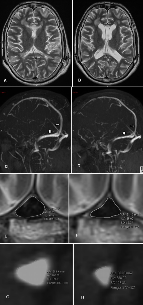
Methods: Twenty patients with chronic hydrocephalus underwent MR venography and MR flow quantification techniques. The venous outflow pressure was estimated from the sinus blood flow and the cross-sectional area of the transverse sinuses. Adult controls as well as a normal young cohort were selected to estimate the change in sinus pressure which occurs throughout life and following the development of hydrocephalus. Significance was tested with a Student's t-test.
Results: The size of the transverse sinuses was unchanged from the 1(st) to the 5(th) decade of life, indicating a stable outflow resistance. However, the blood flow was reduced by 42%, indicating a likely similar reduction in pressure gradient across the sinuses. The sinuses of hydrocephalus patients were 38% smaller than matched controls, indicating a 2.5 times increase in resistance. Despite the 24% reduction in blood flow, a significant increase in sinus pressure is suggested.
Conclusions: The size of the venous sinuses normally does not change over the age range investigated but sinus pressure is reduced proportional to an age-related blood flow reduction. Hydrocephalus is associated with much smaller sinuses than normal and an elevation in venous pressure may explain the lack of CSF absorption into the arachnoid granulations in chronic hydrocephalus.
Keywords: Cerebral blood flow; Chronic hydrocephalus; MR venography; Normal pressure hydrocephalus; Sagittal sinus pressure.
Figures
References
-
- Davson H, Welch K, Segal MB. Physiology and Pathophysiology of the Cerebrospinal Fluid. New York, NY: Churchill Livingstone; 1987. pp. 485–521.
-
- Bateman GA. Hypertensive slit ventricle syndrome: pseudotumor cerebri with a malfunctioning shunt? J Neurosurg. 2013;199:1503–1510. - PubMed
LinkOut - more resources
Full Text Sources
Other Literature Sources
