. 2014:2:693-702.
doi: 10.1039/C3BM60227C.

Incorporation of Sulfated Hyaluronic Acid Macromers into Degradable Hydrogel Scaffolds for Sustained Molecule Delivery

Affiliations

Incorporation of Sulfated Hyaluronic Acid Macromers into Degradable Hydrogel Scaffolds for Sustained Molecule Delivery

Brendan P Purcell et al. Biomater Sci. 2014.

Abstract

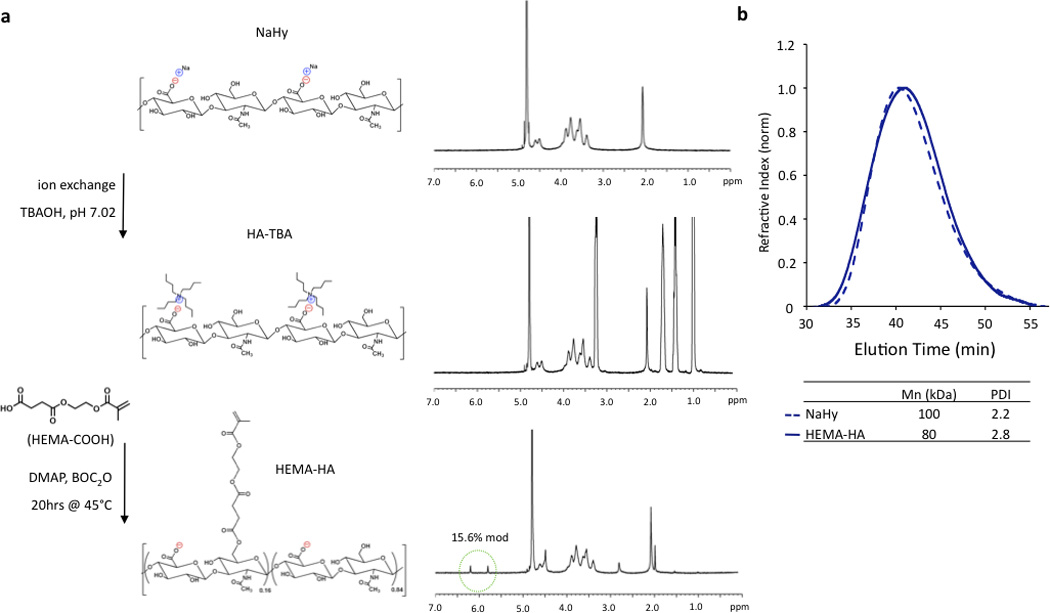
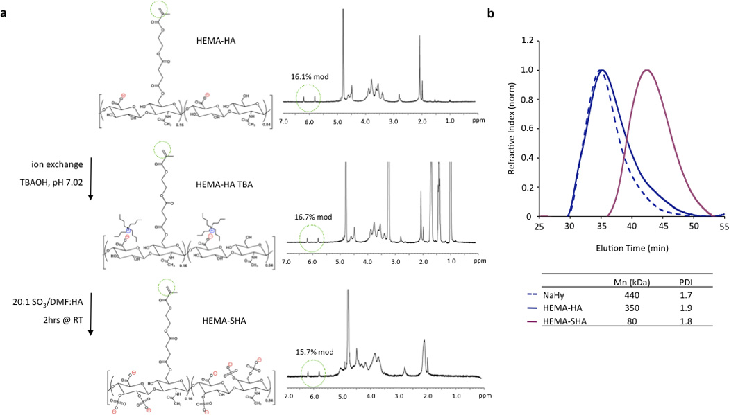
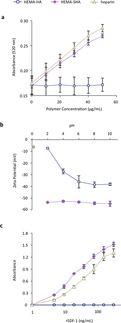
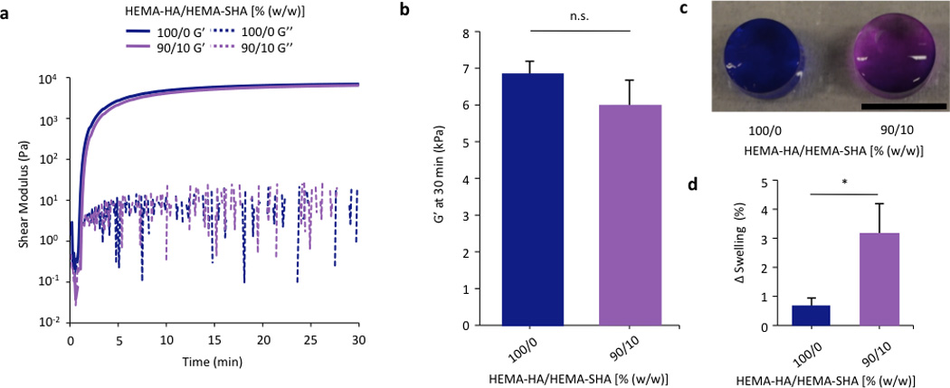
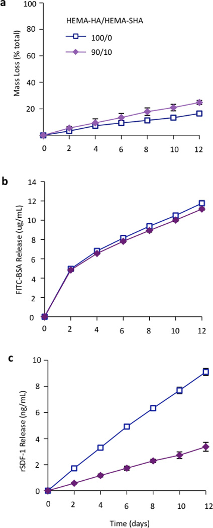
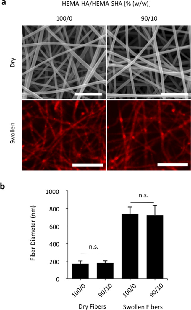
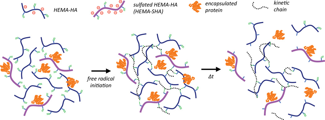
Synthetically sulfated hyaluronic acid (HA) has been shown to bind proteins with high affinity through electrostatic interactions. While HA-based hydrogels have been used widely in recent years for drug delivery and tissue engineering applications, incorporation of sulfated HA into these networks to attenuate the release of proteins has yet to be explored. Here, we developed sulfated and methacrylate-modified HA macromers and incorporated them into HA hydrogels through free radical-initiated crosslinking. The sulfated HA macromers bound a heparin-binding protein (i.e., stromal cell-derived factor 1-α, SDF-1α) with an affinity comparable to heparin and did not alter the gelation behavior or network mechanics when copolymerized into hydrogels at low concentrations. Further, these macromers were incorporated into electrospun nanofibrous hydrogels to introduce sulfate groups into macroporous scaffolds. Once incorporated into either uniform or fibrous HA hydrogels, the sulfated HA macromers significantly slowed encapsulated SDF-1α release over 12 days. Thus, these macromers provide a useful way to introduce heparin-binding features into radically-crosslinked hydrogels to alter protein interactions for a range of applications.


Figures

Figure 1
Figure 1. Synthesis of HEMA modified HA
(a) Sodium hyaluronate (NaHy) was converted to a tertbutylammonium (TBA) salt and reacted with HEMA-succinate in dimethyl sulfoxide using dimethylaminopyridine (DMAP)/di-tert-butyl dicarbonate (BOC2O) catalyzed esterification. HEMA modification was quantified by methacrylate group 1H NMR peaks (green circle). (b) This modification slightly reduced the number average molecular weight (Mn) and increased the polydispersity index (PDI) as measured with GPC when compared to unmodified NaHy.
Figure 2
Figure 2. Synthesis of sulfated HEMA-HA
(a) HEMA-HA was converted to a TBA salt and reacted with SO3/dimethylformamide (DMF) complex in DMF. This reaction altered hydroxyl group 1H NMR peaks as expected, but did not affect methacrylate 1H NMR peaks (green circles). (b) Sulfation reduced the Mn of HEMA-HA by almost 80% as measured by GPC, so a larger 440kDa NaHy was used to generate HEMA-SHA macromers with comparable Mn to HEMA-HA macromers (~80kDa).
Figure 3
Figure 3. Characterization of HEMA-SHA macromers
(a) Sulfate content was increased in HEMA-SHA macromers when compared to HEMA-HA macromers, as determined by dimethylmethylene blue assay. Sulfate content of HEMA-SHA macromers was comparable to that of heparin. (b) Addition of sulfate groups enhanced the negative charge of carboxylic acid-containing HEMA-HA macromers via zeta potential measurements and (c) enhanced binding to the heparin binding protein SDF-1α. rSDF-1α binding of HEMA-SHA macromers was comparable to that of heparin. (n=3 replicates per group, mean±SD).
Figure 4
Figure 4. Crosslinking of HEMA-HA/HEMA-SHA hydrogels
HEMA-HA/HEMA-SHA hydrogels were crosslinked using APS/TEMED free-radical initiators and 4wt% macromers in PBS at 100/0 or 90/10 HEMA-HA/HEMA-SHA compositions (wt/wt). (a) Representative storage (G’) and loss (G’’) moduli profiles over time with and without 10% HEMA-SHA incorporation. (b) Incorporation of 10% HEMA-SHA did not significantly affect G’ after crosslinking had reached a plateau at 30 min. (c) Incorporation of sulfate groups in the hydrogels was visualized using dimethylmethylene blue with purple indicative of sulfate groups (scale bar = 5 mm). (d) Incorporation of sulfate groups slightly increased swelling of the hydrogels after 2 days in PBS. (n=3 replicates per group, mean±SD, *p<0.05 between groups).
Figure 5
Figure 5. Hydrogel degradation and encapsulated protein release
(a) Hydrogel mass loss as determined by uronic acid content. Incorporation of HEMA-SHA macromers enhanced hydrolytic degradation of the hydrogels over 2 weeks. (b) Encapsulated FITC-BSA and (c) heparin-binding SDF-1α release from hydrogels of HEMA-HA alone or incorporating HEMA-SHA macromers. Encapsulated SDF-1α release was reduced by over 50% with incorporation of HEMA-SHA macromers. (n=3 hydrogels per group, mean±SD).
Figure 6
Figure 6. Electrospun HEMA-HA/HEMA-SHA nanofibers
Hydrogels were processed into nanoscale fibers using an electrospinning technique. (a) Fibers visualized in the dry state using scanning electron microscopy (top, scale bars = 2 µm) and after swelling in PBS using fluorescence of incorporated methacrylated rhodamine and optical microscopy (bottom, scale bars = 10 µm). (b) Fiber diameter quantification showed no significant difference between 100/0 and 90/10 HEMA-HA/HEMA-SHA ratios (w/w) in either dry or swollen states (n=80–90 fibers, mean±SD, p>0.05).
Figure 7
Figure 7. Electrostatic interactions in nanofibers
(a) Sulfation of electrospun nanofibers was visualized with dimethylmethylene blue staining. Incorporation of sulfated HEMA-SHA macromers gave the fibers a more negative charge as indicated by the uniform purple stain of the electrospun mat compared to the blue unsulfated HEMA-HA mat (top, scale bar = 10 mm). DMMB staining was localized to individual nanofibers (bottom, scale bars = 50 µm). (b) Incorporation of sulfated HEMA-SHA macromers reduced the release of encapsulated rSDF-1α from the nanofibers by over 50%, indicating stronger electrostatic interactions between the heparin-binding rSDF-1α and sulfated HA (n=3 hydrogels per group, mean±SD).
Schematic 1
Schematic 1. Incorporation of sulfated hyaluronic acid (HA) macromers into hydrogels through free-radical initiated crosslinking
Hydroxyethylmethacrylate (HEMA) modified HA macromers were synthesized with or without sulfate groups to control sulfated polymer concentration in hydrogels by simply blending the two macromers together and crosslinking with free-radical initiators. Release of encapsulated proteins in these networks is controlled by electrostatic interactions with the negatively charged polymers and by gel degradation through ester group hydrolysis of the HEMA crosslinks.

Similar articles

Cited by

References

    1. Burdick JA, Prestwich GD. Adv Mater. 23:H41–H56. - PMC - PubMed
    1. Laurent TC, Fraser JR. FASEB J. 1992;6:2397–2404. - PubMed
    1. Yamada T, Kawasaki T. J Biosci Bioeng. 2005;99:521–528. - PubMed
    1. Guvendiren M, Burdick JA. Nat Commun. 3:792. - PubMed
    1. Khetan S, Guvendiren M, Legant WR, Cohen DM, Chen CS, Burdick JA. Nat Mater. 12:458–465. - PMC - PubMed