Review
. 2014:2014:658243.
doi: 10.1155/2014/658243. Epub 2014 May 18.

Alterations of eye movement control in neurodegenerative movement disorders

Affiliations
Review

Alterations of eye movement control in neurodegenerative movement disorders

Martin Gorges et al. J Ophthalmol. 2014.

Abstract

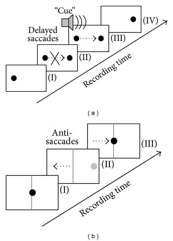
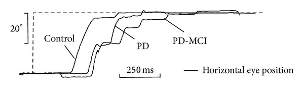
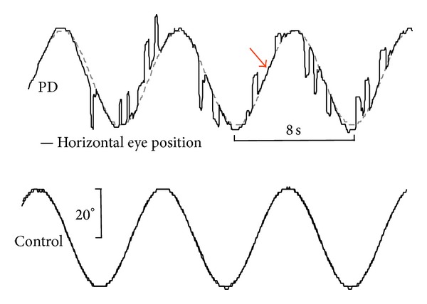
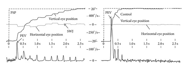
The evolution of the fovea centralis, the most central part of the retina and the area of the highest visual accuracy, requires humans to shift their gaze rapidly (saccades) to bring some object of interest within the visual field onto the fovea. In addition, humans are equipped with the ability to rotate the eye ball continuously in a highly predicting manner (smooth pursuit) to hold a moving target steadily upon the retina. The functional deficits in neurodegenerative movement disorders (e.g., Parkinsonian syndromes) involve the basal ganglia that are critical in all aspects of movement control. Moreover, neocortical structures, the cerebellum, and the midbrain may become affected by the pathological process. A broad spectrum of eye movement alterations may result, comprising smooth pursuit disturbance (e.g., interrupting saccades), saccadic dysfunction (e.g., hypometric saccades), and abnormal attempted fixation (e.g., pathological nystagmus and square wave jerks). On clinical grounds, videooculography is a sensitive noninvasive in vivo technique to classify oculomotion function alterations. Eye movements are a valuable window into the integrity of central nervous system structures and their changes in defined neurodegenerative conditions, that is, the oculomotor nuclei in the brainstem together with their directly activating supranuclear centers and the basal ganglia as well as cortical areas of higher cognitive control of attention.


Figures

Figure 1
Figure 1
Illustration of cognitive demanding oculomotor tasks in order to assess attentional control. (a) In delayed saccades, subjects are asked to fixate the spot (I) and to withhold their gaze shift towards a new randomly appearing target (II) until an acoustic “cue” is sounded (III). The “run” is finished when the old target varnishes (IV). (b) The antisaccade task requires the subject to focus on the center position (I) until a new target (grey) presents randomly at either the right or the left eccentric position (II). The subject is immediately asked to shift the gaze to the contralateral half-plane (away from the target). The “run” is completed by shifting the gaze back onto the central position (III). Black dotted arrows illustrate the required gaze shift.
Figure 2
Figure 2
Visually guided horizontal reflexive saccade in a Parkinson's disease patient with mild cognitive impairment (PD-MCI) and a Parkinson's disease patient without cognitive impairment (PD), compared with an age-matched healthy control subject. Both PD patients presented with a considerable multistep sequence (saccadic hypometria) to shift their gaze onto the target (dashed-line). The videooculographically recorded data display the orthogonalized position for the cyclopean eye shown for the horizontal component (black lines). x-axis: acquisition time in seconds; y-axis: horizontal eye position in degrees.
Figure 3
Figure 3
Horizontal smooth pursuit eye movements (SPEM) elicited by a sinusoidal oscillating spot (f = 0.125 Hz) and exemplified for a Parkinson's disease patient (PD, upper panel) and a representative age-matched healthy control (lower panel). The PD patient presents with severely affected SPEM, frequently interrupted by anticipatory saccades. Although SPEM is heavily impaired in PD, patients retained the ability to perform episodes of genuine smooth pursuit (arrow). For recording details, see Figure 2.
Figure 4
Figure 4
Videooculographic recordings depicting an upward visually guided reflexive saccade elicited in a sudden target jump ranging from −15 to +15 degrees in a patient with progressive supranuclear palsy (PSP, left panel) compared with an age-matched healthy control (right panel). The PSP patient reaches the target (dashed line) in a pathological multistep pattern, approximately 2.5 seconds (x-axis) after new stimulus appearance (black line, vertical gaze position, and y-axis) whereas the control's gaze shift is accomplished after about 600 milliseconds. Abnormal horizontal square wave jerks (SWJ) manifest in the orthogonalized horizontal eye position (gray line in the left panel), together with vertical saccades indicating a curved trajectory. In contrast, the horizontal eye position in the control subject (gray line in the right panel) exhibits no alteration. The lower row shows the corresponding vertical eye velocity (y-axis) computed by use of sample-by-sample differences of the vertical eye position signal. The PSP patient (left) fails in generating larger saccades resulting in a reduced peak eye velocity (PEV) compared with the control subject.

Similar articles

Cited by

References

    1. Goldberg ME, Walker MF. The control of gaze. In: Hudspeth AJ, Schwartz JH, Jessell TM, Siegelbaum SA, Kandel ER, editors. Principles of Neural Science. 5th edition. New York, NY, USA: McGraw-Hill; 2013. pp. 894–916.
    1. Leigh RJ, Zee DS. The Neurology of Eye Movements. 4th edition. New York, NY, USA: Oxford University Press; 2006.
    1. Anderson TJ, MacAskill MR. Eye movements in patients with neurodegenerative disorders. Nature Reviews Neurology. 2013;9(2):74–85. - PubMed
    1. MacAskill MR, Graham CF, Pitcher TL, et al. The influence of motor and cognitive impairment upon visually-guided saccades in Parkinson’s disease. Neuropsychologia. 2012;50(14):3338–3347. - PubMed
    1. Pinkhardt EH, Kassubek J. Ocular motor abnormalities in Parkinsonian syndromes. Parkinsonism & Related Disorders. 2011;17(4):223–230. - PubMed

LinkOut - more resources