How does calcification influence plaque vulnerability? Insights from fatigue analysis
- PMID: 24955401
- PMCID: PMC3997847
- DOI: 10.1155/2014/417324
How does calcification influence plaque vulnerability? Insights from fatigue analysis
Abstract
Background: Calcification is commonly believed to be associated with cardiovascular disease burden. But whether or not the calcifications have a negative effect on plaque vulnerability is still under debate.
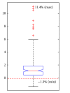
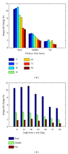
Methods and results: Fatigue rupture analysis and the fatigue life were used to evaluate the rupture risk. An idealized baseline model containing no calcification was first built. Based on the baseline model, we investigated the influence of calcification on rupture path and fatigue life by adding a circular calcification and changing its location within the fibrous cap area. Results show that 84.0% of calcified cases increase the fatigue life up to 11.4%. For rupture paths 10D far from the calcification, the life change is negligible. Calcifications close to lumen increase more fatigue life than those close to the lipid pool. Also, calcifications in the middle area of fibrous cap increase more fatigue life than those in the shoulder area.
Conclusion: Calcifications may play a positive role in the plaque stability. The influence of the calcification only exists in a local area. Calcifications close to lumen may be influenced more than those close to lipid pool. And calcifications in the middle area of fibrous cap are seemly influenced more than those in the shoulder area.
Figures


Similar articles
-
Effect of calcification on the mechanical stability of plaque based on a three-dimensional carotid bifurcation model.BMC Cardiovasc Disord. 2012 Feb 15;12:7. doi: 10.1186/1471-2261-12-7. BMC Cardiovasc Disord. 2012. PMID: 22336469 Free PMC article.
-
Morphometric and Mechanical Analyses of Calcifications and Fibrous Plaque Tissue in Carotid Arteries for Plaque Rupture Risk Assessment.IEEE Trans Biomed Eng. 2021 Apr;68(4):1429-1438. doi: 10.1109/TBME.2020.3038038. Epub 2021 Mar 18. IEEE Trans Biomed Eng. 2021. PMID: 33186100
-
Microcalcifications, Their Genesis, Growth, and Biomechanical Stability in Fibrous Cap Rupture.Adv Exp Med Biol. 2018;1097:129-155. doi: 10.1007/978-3-319-96445-4_7. Adv Exp Med Biol. 2018. PMID: 30315543 Review.
-
Fatigue crack propagation analysis of plaque rupture.J Biomech Eng. 2013 Oct 1;135(10):101003-9. doi: 10.1115/1.4025106. J Biomech Eng. 2013. PMID: 23897295
-
Small entities with large impact: microcalcifications and atherosclerotic plaque vulnerability.Curr Opin Lipidol. 2014 Oct;25(5):327-32. doi: 10.1097/MOL.0000000000000105. Curr Opin Lipidol. 2014. PMID: 25188916 Free PMC article. Review.
Cited by
-
Prognostic impact of intracranial arteriosclerosis subtype after endovascular treatment for acute ischaemic stroke.Eur J Neurol. 2025 Jan;32(1):e16509. doi: 10.1111/ene.16509. Epub 2024 Oct 17. Eur J Neurol. 2025. PMID: 39417311 Free PMC article.
-
Irregular surface of carotid atherosclerotic plaque is associated with ischemic stroke: a magnetic resonance imaging study.J Geriatr Cardiol. 2019 Dec;16(12):872-879. doi: 10.11909/j.issn.1671-5411.2019.12.002. J Geriatr Cardiol. 2019. PMID: 31911791 Free PMC article.
-
Serum Homocysteine and Vascular Calcification: Advances in Mechanisms, Related Diseases, and Nutrition.Korean J Fam Med. 2022 Sep;43(5):277-289. doi: 10.4082/kjfm.21.0227. Epub 2022 Sep 20. Korean J Fam Med. 2022. PMID: 36168899 Free PMC article.
-
Clinical quantitative susceptibility mapping (QSM): Biometal imaging and its emerging roles in patient care.J Magn Reson Imaging. 2017 Oct;46(4):951-971. doi: 10.1002/jmri.25693. Epub 2017 Mar 10. J Magn Reson Imaging. 2017. PMID: 28295954 Free PMC article. Review.
-
FGF-23 levels in patients with critical carotid artery stenosis.Intern Emerg Med. 2015 Jun;10(4):437-44. doi: 10.1007/s11739-014-1183-3. Epub 2015 Jan 9. Intern Emerg Med. 2015. PMID: 25573621
References
-
- Hatsukami TS, Ross R, Polissar NL, Yuan C. Visualization of fibrous cap thickness and rupture in human atherosclerotic carotid plaque in vivo with high-resolution magnetic resonance imaging. Circulation. 2000;102(9):959–964. - PubMed
-
- Finet G, Ohayon J, Rioufol G. Biomechanical interaction between cap thickness, lipid core composition and blood pressure in vulnerable coronary plaque: impact on stability or instability. Coronary Artery Disease. 2004;15(1):13–20. - PubMed
-
- Stary HC. Natural history of calcium deposits in atherosclerosis progression and regression. Zeitschrift fur Kardiologie. 2000;89(supplement 2):28–35. - PubMed
-
- Raggi P, Boulay A, Chasan-Taber S, et al. Cardiac calcification in adult hemodialysis patients: a link between end-stage renal disease and cardiovascular disease? Journal of the American College of Cardiology. 2002;39(4):695–701. - PubMed
-
- Arad Y, Goodman KJ, Roth M, Newstein D, Guerci AD. Coronary calcification, coronary disease risk factors, C-reactive protein, and atherosclerotic cardiovascular disease events: the St. Francis heart study. Journal of the American College of Cardiology. 2005;46(1):158–165. - PubMed
Publication types
MeSH terms
LinkOut - more resources
Full Text Sources
Other Literature Sources
Miscellaneous

