. 2014 Jun 23;9(6):e99818.
doi: 10.1371/journal.pone.0099818. eCollection 2014.

(-)-Phenserine attenuates soman-induced neuropathology

Affiliations

(-)-Phenserine attenuates soman-induced neuropathology

Jun Chen et al. PLoS One. .

Abstract

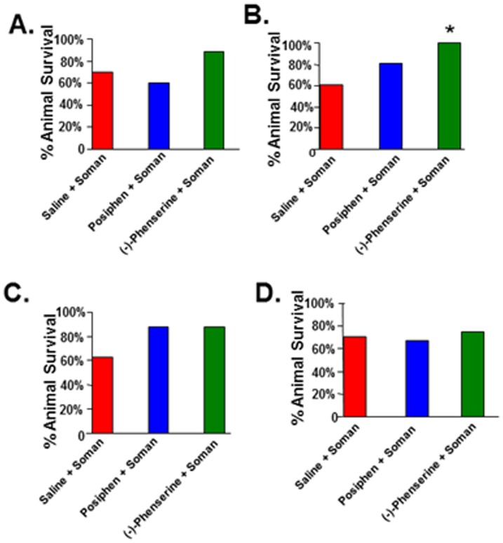
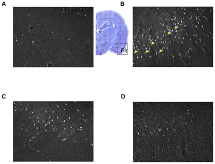
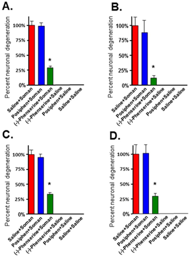
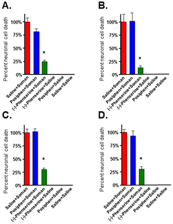
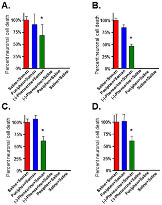
Organophosphorus (OP) nerve agents are deadly chemical weapons that pose an alarming threat to military and civilian populations. The irreversible inhibition of the critical cholinergic degradative enzyme acetylcholinesterase (AChE) by OP nerve agents leads to cholinergic crisis. Resulting excessive synaptic acetylcholine levels leads to status epilepticus that, in turn, results in brain damage. Current countermeasures are only modestly effective in protecting against OP-induced brain damage, supporting interest for evaluation of new ones. (-)-Phenserine is a reversible AChE inhibitor possessing neuroprotective and amyloid precursor protein lowering actions that reached Phase III clinical trials for Alzheimer's Disease where it exhibited a wide safety margin. This compound preferentially enters the CNS and has potential to impede soman binding to the active site of AChE to, thereby, serve in a protective capacity. Herein, we demonstrate that (-)-phenserine protects neurons against soman-induced neuronal cell death in rats when administered either as a pretreatment or post-treatment paradigm, improves motoric movement in soman-exposed animals and reduces mortality when given as a pretreatment. Gene expression analysis, undertaken to elucidate mechanism, showed that (-)-phenserine pretreatment increased select neuroprotective genes and reversed a Homer1 expression elevation induced by soman exposure. These studies suggest that (-)-phenserine warrants further evaluation as an OP nerve agent protective strategy.


Conflict of interest statement

Competing Interests: The authors have declared that no competing interests exist.

Figures

Figure 1
Figure 1. Administration of (−)-phenserine prior to but not after soman protects against soman-induced mortality.
Rats were administered (−)-phenserine, posiphen or saline 4 hr (A), or 30 min (B) prior to or 5 min (C) or 30 min (D) after soman. In the 4 hr pretreatment groups of animals, 12 rats died in the posiphen group and 11 rats died in the control group. There were no deaths in the (−)-phenserine group. In the 30 min pretreatment groups of animals, 2 animals died in the (−)-phenserine group. The bar represents the percent of surviving rats 24 hr after soman exposure calculated as: number of surviving rats 24 hr after soman/total number of rats ×100. n = 17–20. *p<0.026 vs saline/soman by Fisher exact test.
Figure 2
Figure 2. Administration with (−)-phenserine but not posiphen thirty minutes prior to soman exposure reduces neuronal cell death in the vulnerable piriform cortex.
Representative photomicrographs of the piriform cortex stained with Fluorojade C staining (A–D, magnification 200×). Animals were injected with saline (A), a single dose of posiphen (1 mg/kg iv) [C] or (−)-pheserine (1 mg/kg iv) [D] thirty min prior to injection of soman (B). Animals were euthanized 24 hours after soman exposure. The piriform cortex (Pir) is outlined in the coronal section. Fluorojade C-positive degenerating neurons are indicated by the arrowheads. There was no statistically significant difference between groups of animals injected with saline and administration of either posiphen or (−)-phenserine in the absence of soman and are not shown.
Figure 3
Figure 3. Administration of (−)-phenserine 4 hr prior to soman protects against soman-induced neuronal cell death.
Rats were pre-treated with (−)-phenserine, posiphen or saline 4 hr prior to soman. Photographs were acquired from three representative fields in each brain region/animal. The bar represents the average percent neuronal cell death ± SD in the pirform cortex (A), hippocampus (B), basolateral amygdala (C), cingulate cortex (D). The number of fluorescein-positive neurons was counted by an investigator that was blinded to the treatment. n = 6/group. *p<0.001 vs saline/soman by ANOVA+Tukey post hoc analysis.
Figure 4
Figure 4. Administration of (−)-phenserine injected intravenously 30 min prior to soman protects neurons against soman-induced neuronal cell death.
Rats were pretreated with (−)-phenserine, posiphen or saline, as indicated 30 min prior to soman. Slides from each brain region were randomly selected. Photographs were acquired from three representative fields per brain region per animal. The number of fluorescein-positive neurons was counted by an investigator that was blinded to the treatment. The bar represents the average percent neuronal cell death ± SD in the pirform cortex (A), hippocampus (B), basolateral amygdala (C), cingulate cortex (D). n = 6/group. *p<0.001 vs saline/soman by ANOVA+Tukey post hoc analysis.
Figure 5
Figure 5. Administration of (−)-phenserine injected intravenously 5 min after soman protects neurons against soman-induced neuronal cell death.
Rats were post-treated with (−)-phenserine, posiphen, or saline five minutes after soman. Three representative images were acquired from each of the four brain regions per animal. The number of fluorescein-positive neurons was counted by an investigated that was blinded to the treatment. The bar represents the average percent neuronal cell death ± SD in the pirform cortex (A), hippocampus (B), basolateral amygdala (C), cingulate cortex (D). n = 6/group. *p<0.001 vs soman/saline by ANOVA+Tukey post hoc analysis.
Figure 6
Figure 6. Administration of (−)-phenserine injected intravenously 30 min after soman protects neurons against soman-induced neuronal cell death.
Rats were post-treated with (−)-phenserine, posiphen or saline thirty minutes after soman. Images from three representative fields were acquired for each of the four brain regions/animal. The number of fluorescein-positive neurons was counted by an investigator that was blinded to the treatment. The bar represents the average percent neuronal cell death ± SD in the pirform cortex (A), hippocampus (B), basolateral amygdala (C), cingulate cortex (D). n = 6/group. *p<0.001 vs soman/saline by ANOVA+Tukey post hoc analysis.

Similar articles

Cited by

References

    1. Newmark J (2007) Nerve agents. Neurologist 13: 20–32. - PubMed
    1. Shih TM, Duniho SM, McDonough JH (2003) Control of nerve agent-induced seizures is critical for neuroprotection and survival. Toxicol Appl Pharmacol 188: 69–80. - PubMed
    1. Romano JA Jr, King JM (2001) Psychological casualties resulting from chemical and biological weapons. Mil Med 166 12 Suppl: 21–22. - PubMed
    1. Okumura T, Takasu N, Ishimatsu S, Miyanoki S, Mitsuhashi A, et al. (1996) Report on 640 Victims of the Tokyo Subway Sarin Attack. Annals of Emergency Medicine 28: 129–136. - PubMed
    1. Nishiwaki Y, Maekawa K, Ogawa Y, Asukai N, Minami M, et al. (2001) Effects of sarin on the nervous system in rescue team staff members and police officers 3 years after the Tokyo subway sarin attack. Environ Health Perspect 109: 1169–1173. - PMC - PubMed

Publication types

MeSH terms