Multicenter Study
. 2014 Aug;21(4):256-67.
doi: 10.3109/09286586.2014.926940. Epub 2014 Jun 23.

Telemedicine approaches to evaluating acute-phase retinopathy of prematurity: study design

Collaborators
Multicenter Study

Telemedicine approaches to evaluating acute-phase retinopathy of prematurity: study design

Graham E Quinn et al. Ophthalmic Epidemiol. 2014 Aug.

Abstract

Purpose: Detecting sight-threatening retinopathy of prematurity (ROP) relies on a diagnostic examination (DE) performed by an experienced ophthalmologist. An alternative may be a telemedicine system where retinal images of at-risk infants are graded by readers to determine features of ROP indicating the need for a DE.

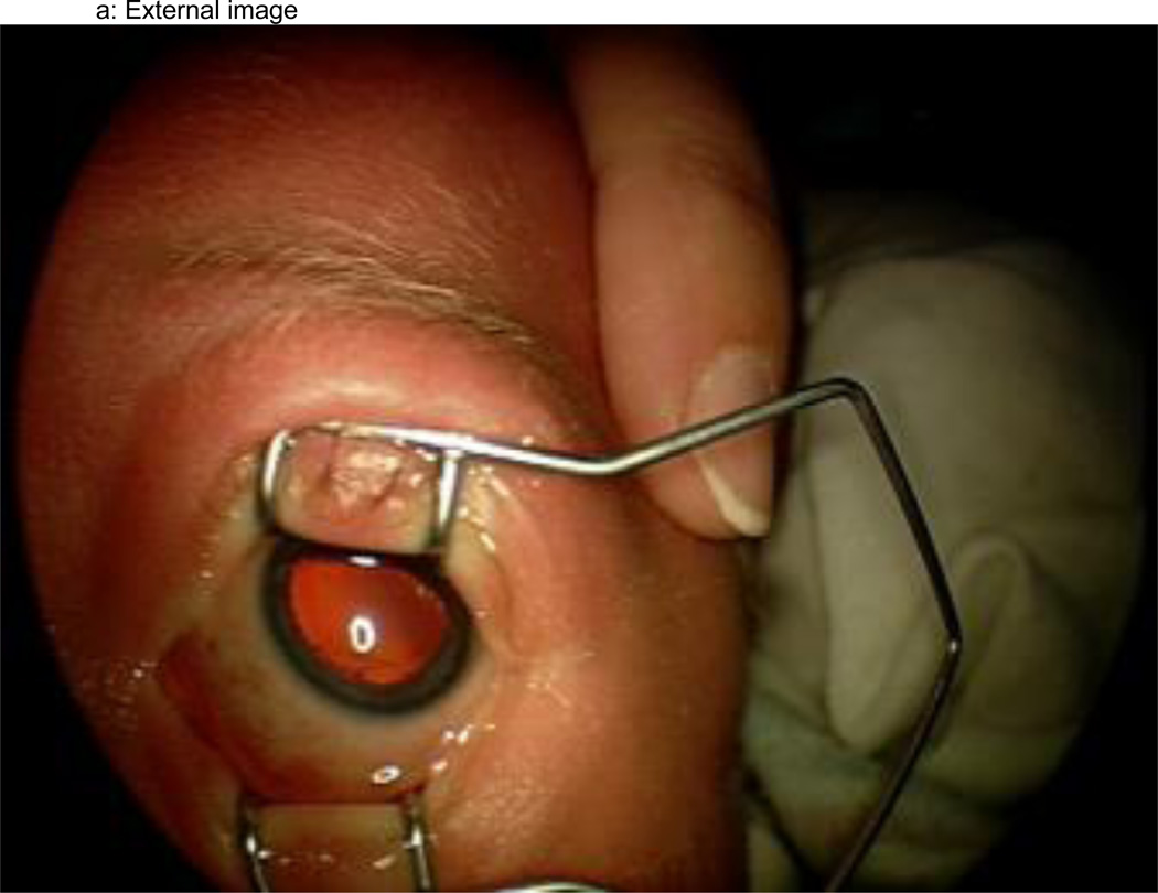
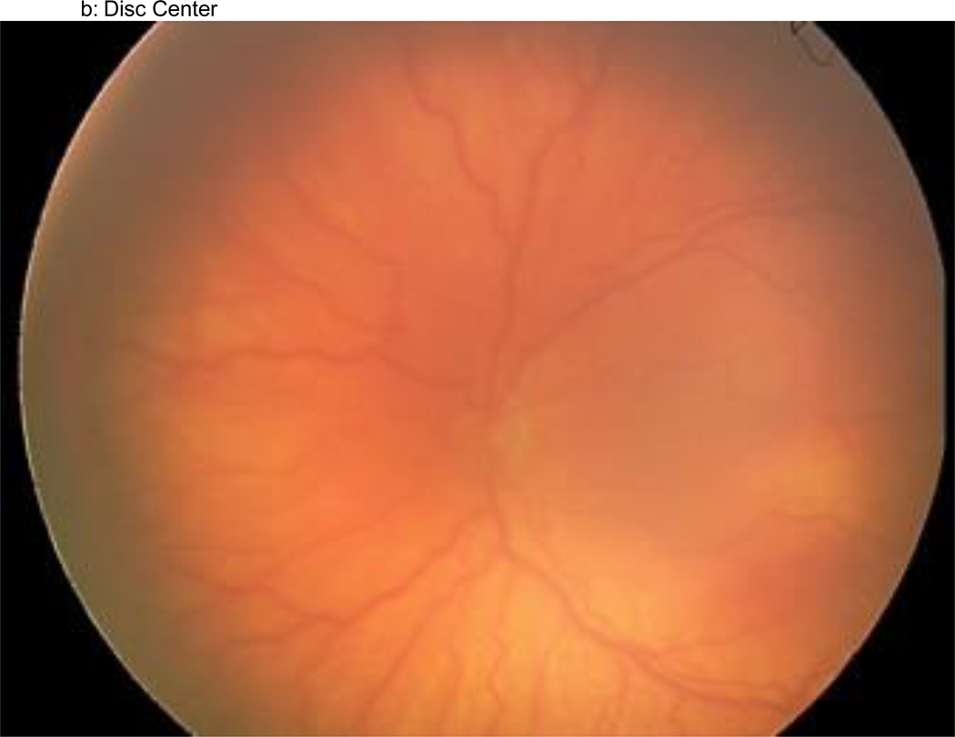
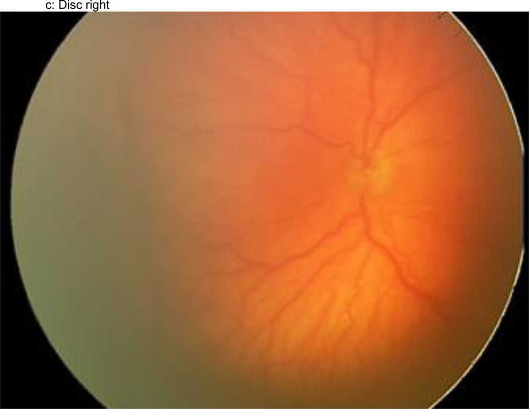
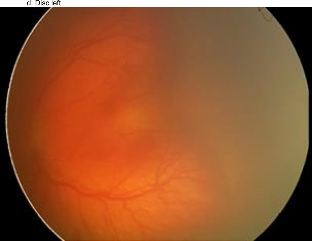
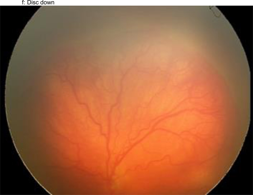
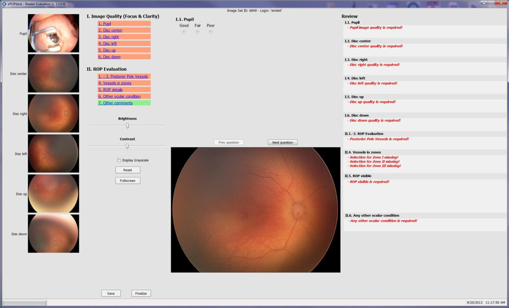
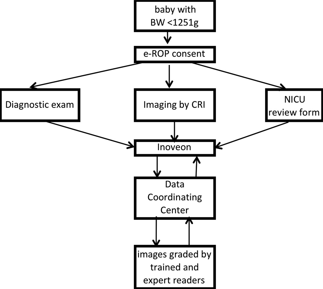
Methods: The multicenter Telemedicine Approaches to Evaluating Acute-phase ROP (e-ROP) Study is a cohort study of 2000 infants with birth weights <1251 g. At each visit, ophthalmologists perform DEs and non-physician imagers obtain iris and five retinal images with the disc positioned in the center, right, left, up and down. Images are uploaded to a secure server for grading by non-physician readers for the detection of plus disease, stage 3 ROP and/or zone I disease, any of which indicates "referral-warranted ROP" (RW-ROP). Images from all infants with RW-ROP and a random sample of infants without RW-ROP (based on DEs) are selected for grading. Gradings are compared to DEs to determine the validity and evaluate reliability, feasibility, safety, and cost-effectiveness of the telemedicine system.

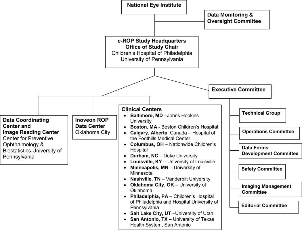
Results: e-ROP is conducted in 12 Clinical Centers in the US and Canada with Study Headquarters, the Data Coordinating Center and the Image Reading Center in Philadelphia and the ROP Data Center in Oklahoma City. A total of 27 study center coordinators, 34 ophthalmologists, 26 imagers, and 4 readers have been certified. All study data are submitted using a secure web-based system.

Conclusion: The design and findings of this study will be useful to conduct other ROP studies or evaluate telemedicine for other diseases.

Trial registration: ClinicalTrials.gov NCT01264276.

Keywords: Childhood blindness; prematurity; retinopathy of prematurity; telemedicine.


Conflict of interest statement

None of the authors have any proprietary interests or conflicts of interest related to this submission.

Figures

Figure 1
Figure 1
a–f: An example of images collected at each imaging session by the trained retinal imager. The standard image set was collected for each eye of the baby and consisted of an image of the anterior segment (Figure 1a) and five retinal fields where the image had the: (1) disc central (Figure 1b), (2) disc right (Figure 1c), (3) disc left (Figure 1d), (4) disc up (Figure 1e), and (5) disc down (Figure 1f). This image set demonstrates the presence of plus disease, stage 3 ROP, and zone I ROP – any one of which would qualify the eye as having RW-ROP by a reader.
Figure 1
Figure 1
a–f: An example of images collected at each imaging session by the trained retinal imager. The standard image set was collected for each eye of the baby and consisted of an image of the anterior segment (Figure 1a) and five retinal fields where the image had the: (1) disc central (Figure 1b), (2) disc right (Figure 1c), (3) disc left (Figure 1d), (4) disc up (Figure 1e), and (5) disc down (Figure 1f). This image set demonstrates the presence of plus disease, stage 3 ROP, and zone I ROP – any one of which would qualify the eye as having RW-ROP by a reader.
Figure 2
Figure 2
The components of the e-ROP Study.
Figure 3
Figure 3
The web-based grading form completed by all Trained and Expert Readers in the e-ROP study.
Figure 4
Figure 4
The workflow in the e-ROP Study from identifying eligible babies to activities conducted at the clinical centers to uploading of Study data to the remote server and subsequent grading of images and analysis of the Study results.

References

    1. Gilbert C, Fielder A, Gordillo L, Quinn G, Semiglia R, Visintin P, Zin A on behalf of the International NO-ROP Group. Characteristics of infants with severe retinopathy of prematurity in countries with low, moderate and high levels of development: Implications for screening programs. Pediatrics. 2005;115:e518–e525. - PubMed
    1. Gilbert C. Retinopathy of prematurity: A global perspective of the epidemics, population of babies at risk and implications for control. Early Human Development. 2008;84:77–82. - PubMed
    1. Kemper AR, Wallace DK. Neonatologists' practices and experiences in arranging retinopathy of prematurity screening services. Pediatrics. 2007;120(3):527–531. - PMC - PubMed
    1. Kemper AR, Freedman SF, Wallace DK. Retinopathy of prematurity care: patterns of care and workforce analysis. J AAPOS. 2008;12(4):344–348. - PMC - PubMed
    1. Castillo-Riquelme MC, Lord J, Moseley MJ, Fielder AR, Haines L. Cost-effectiveness of digital photographic screening for retinopathy of prematurity in the United Kingdom. Int J Technol Assess Health Care. 2004;20(2):201–213. - PubMed

Publication types

Associated data