. 2014 Jun 23;9(6):e100481.
doi: 10.1371/journal.pone.0100481. eCollection 2014.

CRISPLD2 is a target of progesterone receptor and its expression is decreased in women with endometriosis

Affiliations

CRISPLD2 is a target of progesterone receptor and its expression is decreased in women with endometriosis

Jung-Yoon Yoo et al. PLoS One. .

Abstract

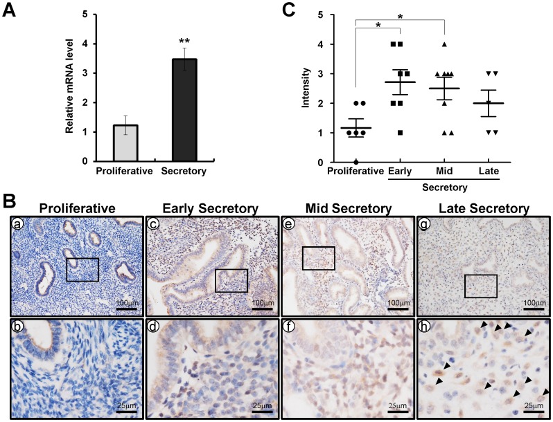
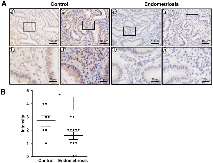
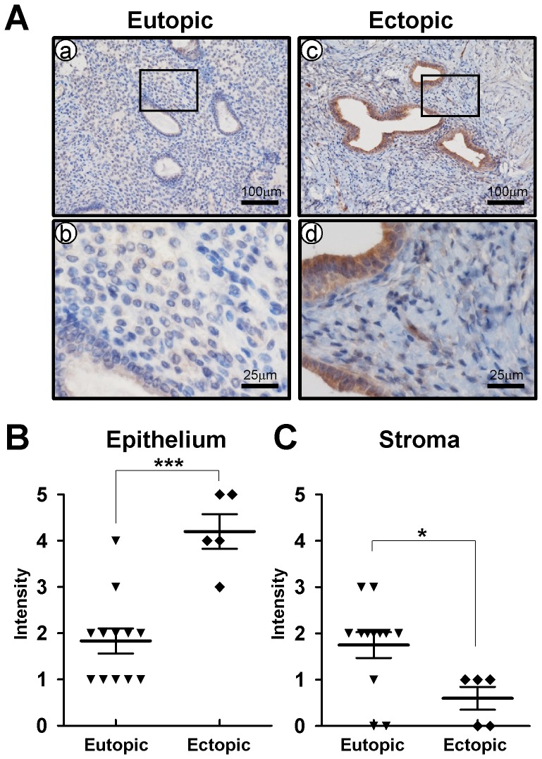
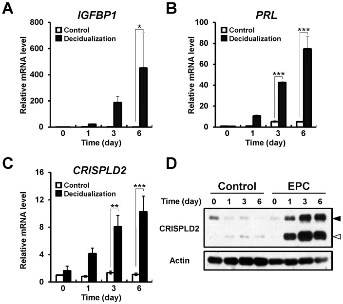
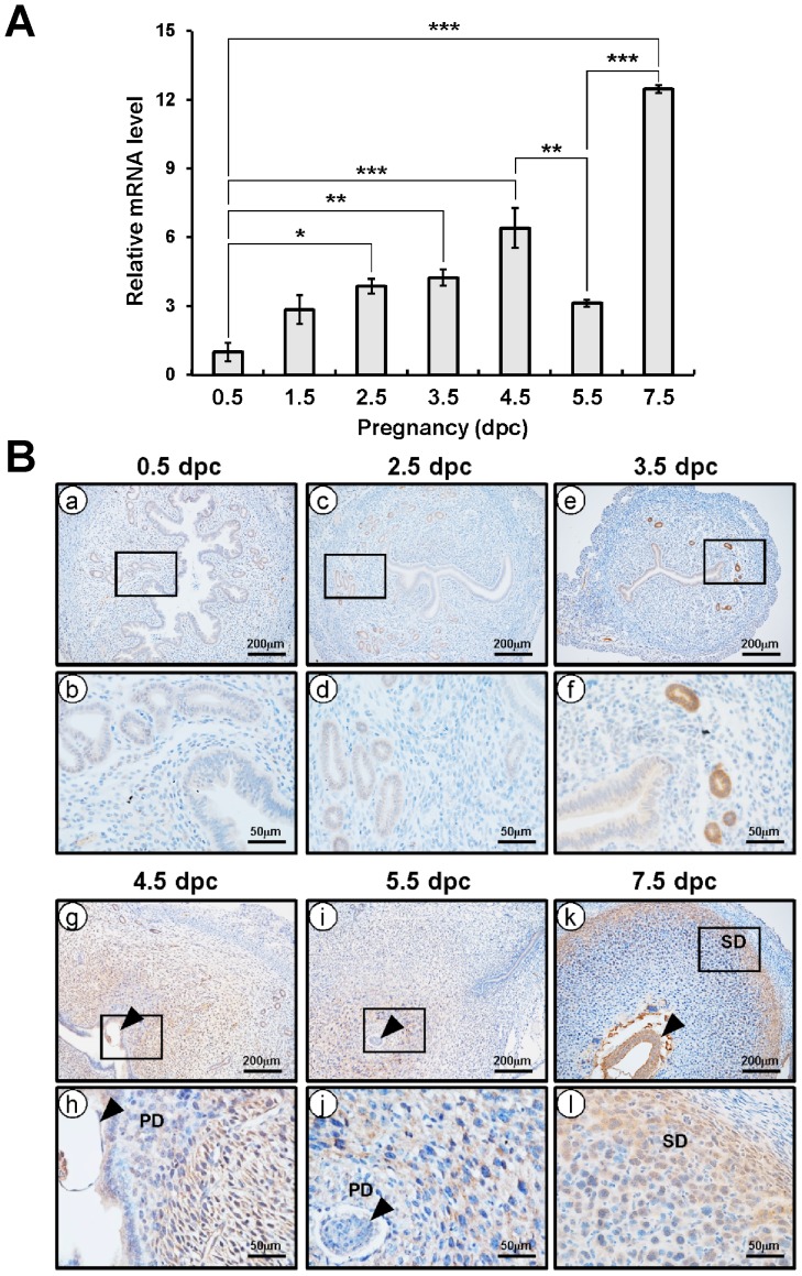
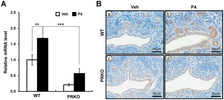
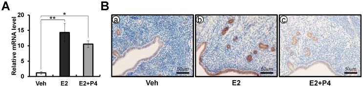
Endometriosis, defined as the presence of endometrial cells outside of the uterine cavity, is a major cause of infertility and pelvic pain, afflicting more than 10% of reproductive age women. Endometriosis is a chronic inflammatory disease and lipopolysaccharide promotes the proliferation and invasion of endometriotic stromal cells. Cysteine-rich secretory protein LCCL domain-containing 2 (CRISPLD2) has high affinity for lipopolysaccharide and plays a critical role in defense against endotoxin shock. However, the function of CRISPLD2 has not been studied in endometriosis and uterine biology. Herein, we examined the expression of CRISPLD2 in endometrium from patients with and without endometriosis using immunohistochemistry. The expression of CRISPLD2 was higher in the secretory phase in human menstrual cycle compared to proliferative phase. The expression of CRISPLD2 was significantly decreased in the endometrium of women with endometriosis in the early secretory phase compared to women without endometriosis. The increase of CRISPLD2 expression at the early secretory and dysregulation of its expression in endometriosis suggest progesterone (P4) regulation of CRISPLD2. To investigate whether CRISPLD2 is regulated by P4, we examined the expression of the CRISPLD2 in the uteri of wild-type and progesterone receptor knock out (PRKO) mice. The expression of CRISPLD2 was significantly increased after P4 treatment in the wild-type mice. However, CRISPLD2 expression was significantly decreased in the (PRKO) mice treated with P4. During early pregnancy, the expression of CRISPLD2 was increased in decidua of implantation and post-implantation stages. CRISPLD2 levels were also increased in cultured human endometrial stromal cells during in vitro decidualization. These results suggest that the CRISPLD2 is a target of the progesterone receptor and may play an important role in pathogenesis of endometriosis.


Conflict of interest statement

Competing Interests: Co-author Jae-wook Jeong is a PLOS ONE Editorial Board member. This does not alter the authors' adherence to PLOS ONE Editorial policies and criteria.

Figures

Figure 1
Figure 1. Comparison of CRISPLD2 expression in the endometrium during menstrual cycle.
(A) Quantitative real-time PCR analysis of CRISPLD2 gene expression in proliferative and secretory phase during the menstrual cycle normalized using the ddCt method to the 18S gene. The results represent the mean ± SEM. ** p<0.01. (B) Immunohistochemical analysis of CRISPLD2 in proliferative (a and b) and early (c and d), mid (e and f), and late (g and h) secretory phase of the menstrual cycle. Black arrow head indicates a decidualized cell. (C) CRISPLD2 was scored by measuring expression intensity of endometrial cells. The results represent the mean ± SEM. * p<0.05.
Figure 2
Figure 2. Comparison of CRISPLD2 expression in the endometrium between women with and without diagnosed endometriosis.
(A) Immunohistochemical analysis of CRISPLD2 in paired (a - d from women without diagnosed endometriosis; e - h from women with endometriosis) endometrium. (B) CRISPLD2 was scored by measuring expression intensity of endometrial cells. The results represent the mean ± SEM. * p<0.05
Figure 3
Figure 3. Comparison of CRISPLD2 expression in the endometrium between eutopic and ectopic endometriosis lesions.
(A) Representative immunohistochemistry of CRISPLD2 in eutopic and ectopic endometrium with endometriosis. (B and C) CRISPLD2 was scored by measuring expression intensity of endometrial epithelial (B) and stromal (C) cells. The results represent the mean ± SEM. *** p<0.001; * p<0.05
Figure 4
Figure 4. Expression of CRISPLD2 during in vitro decidualization of hESCs.
(A to C) Expression of decidualization marker genes, IGFPB1 (A) and PRL (B) and CRISPLD2 gene (C) were examined during in vitro decidualization of hESCs. The results represent the mean ± SEM. * p<0.05; **, p<0.01; ***, p<0.001. (D) Protein level of CRISPLD2 was measured by Western blot analysis. Actin was used as loading control. Black arrow head indicates a non-secreted CRISPLD2 and white arrow head indicates secreted CRISPLD2.
Figure 5
Figure 5. The expression level of Crispld2 in pregnancy and the localization pattern of CRISPLD2 during early pregnancy.
(A) The expression level of Crispld2 was measured in uteri of early pregnancy. Total RNA used for the quantitative real-time PCR assays was prepared from early pregnancy uteri. The results represent the mean ± SEM of three independent RNA sets. * p<0.05; ** p<0.01; ***; p<0.001. (B) The localization pattern of CRISPLD2 during natural pregnancy by immunohistochemistry were determined at 0.5 dpc (a and b), 2.5 dpc (c and d), 3.5 dpc (e and f), 4.5 dpc (g and h), 5.5 dpc (i and j), and 7.5 dpc (k and l). Black arrow head indicates embryo, PD means primary differentiated decidual cells, and SD means secondary decidual zone.
Figure 6
Figure 6. The expression of CRISPLD2 in wild-type or PRKO mice.
(A) The expression level of Crispld2 from P4 treated wild-type or PRKO uteri by quantitative real-time PCR. Total RNA used for the quantitative real-time PCR was prepared from wild-type or PRKO uteri treated with P4 or vehicle for 6 hours. The results represent the mean ± SEM of three independent RNA sets. ** p<0.01; *** p<0.001. (B) The localization pattern of CRISPLD2 by immunohistochemistry in vehicle or P4-treated uteri. Uterine sections were collected from P4 or Veh treated wild-type (a and b) and PRKO (c and d) mice for 6 hours.
Figure 7
Figure 7. The expression of CRISPLD2 by steroid hormones.
(A) The expression level of CRISPLD2 from vehicle, or estrogen (E2) treated uteri by real-time PCR. Total RNA used for the real-time PCR assays was prepared from ovariectomized wild-type C57BL/6 mice treated with vehicle, E2, or E2+P4 for 6 hours. The results represent the mean ± SEM of three independent RNA sets. * p<0.5; ** p<0.01. (B) The localization pattern of CRISPLD2 by immunohistochemistry in vehicle, E2, or E2+P4 treated wild-type mice uteri for 6 hours. Nuclei were counterstained with hematoxylin.

References

    1. Eskenazi B, Warner ML (1997) Epidemiology of endometriosis. Obstet Gynecol Clin North Am 24: 235–258. - PubMed
    1. Bulun SE (2009) Endometriosis. N Engl J Med 360: 268–279. - PubMed
    1. Sinaii N, Cleary SD, Ballweg ML, Nieman LK, Stratton P (2002) High rates of autoimmune and endocrine disorders, fibromyalgia, chronic fatigue syndrome and atopic diseases among women with endometriosis: a survey analysis. Hum Reprod 17: 2715–2724. - PubMed
    1. Olive DL, Schwartz LB (1993) Endometriosis. N Engl J Med 328: 1759–1769. - PubMed
    1. Kao LC, Germeyer A, Tulac S, Lobo S, Yang JP, et al. (2003) Expression profiling of endometrium from women with endometriosis reveals candidate genes for disease-based implantation failure and infertility. Endocrinology 144: 2870–2881. - PubMed

Publication types

MeSH terms